 Movie
Movie Controller

[English] 日本語
 Yorodumi
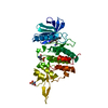
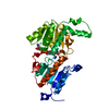
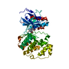
- PDB-7dg4: The co-crystal structure of DYRK2 with a small molecule inhibitor 6 -
+ Open data

- Basic information
| Entry | Database: PDB / ID: 7dg4 | ||||||
|---|---|---|---|---|---|---|---|
| Title | The co-crystal structure of DYRK2 with a small molecule inhibitor 6 | ||||||
 Components Components | Dual specificity tyrosine-phosphorylation-regulated kinase 2 | ||||||
 Keywords Keywords | CELL CYCLE / kinase / inhibitor | ||||||
| Function / homology |  Function and homology information Function and homology informationdual-specificity kinase / negative regulation of calcineurin-NFAT signaling cascade / smoothened signaling pathway / intrinsic apoptotic signaling pathway in response to DNA damage by p53 class mediator / positive regulation of glycogen biosynthetic process / ubiquitin ligase complex / protein serine/threonine/tyrosine kinase activity / regulation of signal transduction by p53 class mediator / manganese ion binding / protein tyrosine kinase activity ...dual-specificity kinase / negative regulation of calcineurin-NFAT signaling cascade / smoothened signaling pathway / intrinsic apoptotic signaling pathway in response to DNA damage by p53 class mediator / positive regulation of glycogen biosynthetic process / ubiquitin ligase complex / protein serine/threonine/tyrosine kinase activity / regulation of signal transduction by p53 class mediator / manganese ion binding / protein tyrosine kinase activity / Regulation of TP53 Activity through Phosphorylation / cytoskeleton / protein phosphorylation / ribonucleoprotein complex / protein serine kinase activity / protein serine/threonine kinase activity / DNA damage response / magnesium ion binding / nucleoplasm / ATP binding / nucleus / cytosol / cytoplasm Similarity search - Function | ||||||
| Biological species |  Homo sapiens (human) Homo sapiens (human) | ||||||
| Method |  X-RAY DIFFRACTION / SYNCHROTRON / MOLECULAR REPLACEMENT / Resolution: 2.58 Å X-RAY DIFFRACTION / SYNCHROTRON / MOLECULAR REPLACEMENT / Resolution: 2.58 Å | ||||||
 Authors Authors | Wei, T. / Xiao, J. | ||||||
| Funding support | 1items
| ||||||
 Citation Citation | Journal: Elife / Year: 2022 Title: Selective inhibition reveals the regulatory function of DYRK2 in protein synthesis and calcium entry. Authors: Wei, T. / Wang, J. / Liang, R. / Chen, W. / Chen, Y. / Ma, M. / He, A. / Du, Y. / Zhou, W. / Zhang, Z. / Zeng, X. / Wang, C. / Lu, J. / Guo, X. / Chen, X.W. / Wang, Y. / Tian, R. / Xiao, J. / Lei, X. | ||||||
| History |
|
- Structure visualization
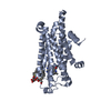
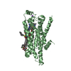
| Structure viewer | Molecule: MolmilJmol/JSmol |
|---|
- Downloads & links
-Download
| PDBx/mmCIF format | 7dg4.cif.gz | 81.9 KB | Display | PDBx/mmCIF format |
|---|---|---|---|---|
| PDB format | pdb7dg4.ent.gz | 59.4 KB | Display | PDB format |
| PDBx/mmJSON format | 7dg4.json.gz | Tree view | PDBx/mmJSON format | |
| Others |  Other downloads Other downloads |
-Validation report
| Arichive directory | https://data.pdbj.org/pub/pdb/validation_reports/dg/7dg4ftp://data.pdbj.org/pub/pdb/validation_reports/dg/7dg4 | HTTPS FTP |
|---|
-Related structure data
| Related structure data | 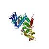 7dh9C 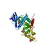 7dhcC  7dhhC 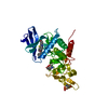 7dhkC  7dhnC 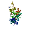 7dhoC 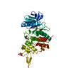 7dhvC 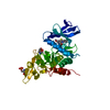 7djoC  7dl6C 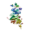 3k2lS S: Starting model for refinement C: citing same article ( |
|---|---|
| Similar structure data |


-Links
 PDBj
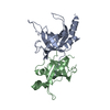
PDBj- Assembly
| Deposited unit | 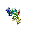
| ||||||||
|---|---|---|---|---|---|---|---|---|---|
| 1 |
| ||||||||
| Unit cell |
| ||||||||
| Components on special symmetry positions |
|
-Components
| #1: Protein | Mass: 37675.633 Da / Num. of mol.: 1 Source method: isolated from a genetically manipulated source Source: (gene. exp.) Homo sapiens (human) / Gene: DYRK2 / Production host:  |
|---|---|
| #2: Chemical | ChemComp-H5R /   Mass: 368.492 Da / Num. of mol.: 1 / Source method: obtained synthetically / Formula: C21H24N2O2S / Feature type: SUBJECT OF INVESTIGATION Mass: 368.492 Da / Num. of mol.: 1 / Source method: obtained synthetically / Formula: C21H24N2O2S / Feature type: SUBJECT OF INVESTIGATION |
| #3: Water | ChemComp-HOH /  Mass: 18.015 Da / Num. of mol.: 23 / Source method: isolated from a natural source / Formula: H2O Mass: 18.015 Da / Num. of mol.: 23 / Source method: isolated from a natural source / Formula: H2O |
| Has ligand of interest | Y |
| Has protein modification | Y |
-Experimental details
-Experiment
| Experiment | Method: X-RAY DIFFRACTION / Number of used crystals: 1 |
|---|
- Sample preparation
| Crystal | Density Matthews: 3.65 Å3/Da / Density % sol: 66.29 % |
|---|---|
| Crystal grow | Temperature: 291 K / Method: vapor diffusion, sitting drop Details: 0.36M-0.5M sodium citrate tribasic dehydrate, 0.01M sodium borate, pH 7.5-9.5 |
-Data collection
| Diffraction | Mean temperature: 293 K / Serial crystal experiment: N |
|---|---|
| Diffraction source | Source: SYNCHROTRON / Site: SSRF  / Beamline: BL17U1 / Wavelength: 1 Å / Beamline: BL17U1 / Wavelength: 1 Å |
| Detector | Type: DECTRIS EIGER X 16M / Detector: PIXEL / Date: Oct 9, 2018 |
| Radiation | Protocol: SINGLE WAVELENGTH / Monochromatic (M) / Laue (L): M / Scattering type: x-ray |
| Radiation wavelength | Wavelength: 1 Å / Relative weight: 1 |
| Reflection | Resolution: 2.41→64.44 Å / Num. obs: 21752 / % possible obs: 99.9 % / Redundancy: 7.9 % / CC1/2: 0.99 / Net I/σ(I): 7.9 |
| Reflection shell | Resolution: 2.41→2.55 Å / Rmerge(I) obs: 0.945 / Num. unique obs: 3121 / CC1/2: 0.818 |
- Processing
| Software |
| ||||||||||||||||||||||||||||||||||||||||||||||||||||||||||||||||||||||||||||||
|---|---|---|---|---|---|---|---|---|---|---|---|---|---|---|---|---|---|---|---|---|---|---|---|---|---|---|---|---|---|---|---|---|---|---|---|---|---|---|---|---|---|---|---|---|---|---|---|---|---|---|---|---|---|---|---|---|---|---|---|---|---|---|---|---|---|---|---|---|---|---|---|---|---|---|---|---|---|---|---|
| Refinement | Method to determine structure: MOLECULAR REPLACEMENT Starting model: 3K2L Resolution: 2.58→64.44 Å / SU ML: 0.39 / Cross valid method: THROUGHOUT / σ(F): 1.37 / Phase error: 27.21 / Stereochemistry target values: ML
| ||||||||||||||||||||||||||||||||||||||||||||||||||||||||||||||||||||||||||||||
| Solvent computation | Shrinkage radii: 0.9 Å / VDW probe radii: 1.11 Å / Solvent model: FLAT BULK SOLVENT MODEL | ||||||||||||||||||||||||||||||||||||||||||||||||||||||||||||||||||||||||||||||
| Displacement parameters | Biso max: 126.81 Å2 / Biso mean: 65.3031 Å2 / Biso min: 30 Å2 | ||||||||||||||||||||||||||||||||||||||||||||||||||||||||||||||||||||||||||||||
| Refinement step | Cycle: final / Resolution: 2.58→64.44 Å
| ||||||||||||||||||||||||||||||||||||||||||||||||||||||||||||||||||||||||||||||
| LS refinement shell | Refine-ID: X-RAY DIFFRACTION / Rfactor Rfree error: 0
|
