 Movie
Movie Controller

[English] 日本語
 Yorodumi
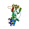
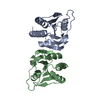
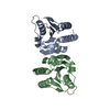
- PDB-7dfx: Structure and function of Diadenylate cyclase DacM in Mycoplasma ... -
+ Open data

- Basic information
| Entry | Database: PDB / ID: 7dfx | ||||||
|---|---|---|---|---|---|---|---|
| Title | Structure and function of Diadenylate cyclase DacM in Mycoplasma ovipneumoniae | ||||||
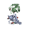
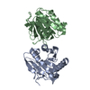
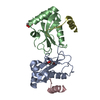
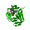
 Components Components | DAC domain-containing protein | ||||||
 Keywords Keywords | IMMUNE SYSTEM / second messenger / c-di-AMP / diadenylate cyclase / Mycoplasma ovipneumoniae | ||||||
| Function / homology |  Function and homology information Function and homology informationdiadenylate cyclase activity / adenylate cyclase activity / ATP binding / membrane Similarity search - Function | ||||||
| Biological species |  Mycoplasma ovipneumoniae 14811 (bacteria) Mycoplasma ovipneumoniae 14811 (bacteria) | ||||||
| Method |  X-RAY DIFFRACTION / SYNCHROTRON / MOLECULAR REPLACEMENT / molecular replacement / Resolution: 2.996 Å X-RAY DIFFRACTION / SYNCHROTRON / MOLECULAR REPLACEMENT / molecular replacement / Resolution: 2.996 Å | ||||||
 Authors Authors | Fan, S. / Li, M. | ||||||
 Citation Citation | Journal: To Be Published Title: Structure and function of Diadenylate cyclase DacM in Mycoplasma ovipneumoniae Authors: Li, M. / Fan, S. | ||||||
| History |
|
- Structure visualization
| Structure viewer | Molecule: MolmilJmol/JSmol |
|---|
- Downloads & links
-Download
| PDBx/mmCIF format | 7dfx.cif.gz | 76.6 KB | Display | PDBx/mmCIF format |
|---|---|---|---|---|
| PDB format | pdb7dfx.ent.gz | 57.4 KB | Display | PDB format |
| PDBx/mmJSON format | 7dfx.json.gz | Tree view | PDBx/mmJSON format | |
| Others |  Other downloads Other downloads |
-Validation report
| Arichive directory | https://data.pdbj.org/pub/pdb/validation_reports/df/7dfxftp://data.pdbj.org/pub/pdb/validation_reports/df/7dfx | HTTPS FTP |
|---|
-Related structure data
| Related structure data | 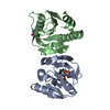 7dg0C  4rv7S S: Starting model for refinement C: citing same article ( |
|---|---|
| Similar structure data |


-Links
 PDBj
PDBj
- Assembly
| Deposited unit | 
| ||||||||
|---|---|---|---|---|---|---|---|---|---|
| 1 |
| ||||||||
| Unit cell |
| ||||||||
| Components on special symmetry positions |
|
-Components
| #1: Protein | Mass: 19507.145 Da / Num. of mol.: 2 Source method: isolated from a genetically manipulated source Source: (gene. exp.) Mycoplasma ovipneumoniae 14811 (bacteria)Gene: MOVI_0530 / Production host:  Escherichia phage EcSzw-2 (virus) / References: UniProt: A0A014M399 Escherichia phage EcSzw-2 (virus) / References: UniProt: A0A014M399#2: Chemical | ChemComp-MLA /   Mass: 104.061 Da / Num. of mol.: 5 / Source method: obtained synthetically / Formula: C3H4O4 / Feature type: SUBJECT OF INVESTIGATION Mass: 104.061 Da / Num. of mol.: 5 / Source method: obtained synthetically / Formula: C3H4O4 / Feature type: SUBJECT OF INVESTIGATION#3: Water | ChemComp-HOH / |  Mass: 18.015 Da / Num. of mol.: 85 / Source method: isolated from a natural source / Formula: H2O Mass: 18.015 Da / Num. of mol.: 85 / Source method: isolated from a natural source / Formula: H2OHas ligand of interest | Y | Has protein modification | N | |
|---|
-Experimental details
-Experiment
| Experiment | Method: X-RAY DIFFRACTION / Number of used crystals: 1 |
|---|
- Sample preparation
| Crystal | Density Matthews: 7.54 Å3/Da / Density % sol: 83.69 % |
|---|---|
| Crystal grow | Temperature: 288 K / Method: vapor diffusion, hanging drop Details: 1 M sodium malonate pH 5.0, 0.1 M citric acid pH 3.5 |
-Data collection
| Diffraction | Mean temperature: 100 K / Serial crystal experiment: N |
|---|---|
| Diffraction source | Source: SYNCHROTRON / Site: SSRF  / Beamline: BL17U / Wavelength: 0.9792 Å / Beamline: BL17U / Wavelength: 0.9792 Å |
| Detector | Type: DECTRIS EIGER X 16M / Detector: PIXEL / Date: Nov 21, 2019 |
| Radiation | Protocol: SINGLE WAVELENGTH / Monochromatic (M) / Laue (L): M / Scattering type: x-ray |
| Radiation wavelength | Wavelength: 0.9792 Å / Relative weight: 1 |
| Reflection | Resolution: 2.99→50 Å / Num. obs: 23720 / % possible obs: 100 % / Redundancy: 36.1 % / CC1/2: 0.999 / Rmerge(I) obs: 0.208 / Net I/σ(I): 23 |
| Reflection shell | Resolution: 3→50 Å / Rmerge(I) obs: 0.208 / Num. unique obs: 23720 / CC1/2: 0.361 |
-Phasing
| Phasing | Method: molecular replacement |
|---|
- Processing
| Software |
| ||||||||||||||||||||||||||||||||||||||||||||||||||||||||||||
|---|---|---|---|---|---|---|---|---|---|---|---|---|---|---|---|---|---|---|---|---|---|---|---|---|---|---|---|---|---|---|---|---|---|---|---|---|---|---|---|---|---|---|---|---|---|---|---|---|---|---|---|---|---|---|---|---|---|---|---|---|---|
| Refinement | Method to determine structure: MOLECULAR REPLACEMENT Starting model: 4rv7 Resolution: 2.996→32.941 Å / SU ML: 0.29 / Cross valid method: THROUGHOUT / σ(F): 1.35 / Phase error: 19.43 / Stereochemistry target values: ML
| ||||||||||||||||||||||||||||||||||||||||||||||||||||||||||||
| Solvent computation | Shrinkage radii: 0.9 Å / VDW probe radii: 1.11 Å / Solvent model: FLAT BULK SOLVENT MODEL | ||||||||||||||||||||||||||||||||||||||||||||||||||||||||||||
| Displacement parameters | Biso max: 141.79 Å2 / Biso mean: 54.4221 Å2 / Biso min: 18.84 Å2 | ||||||||||||||||||||||||||||||||||||||||||||||||||||||||||||
| Refinement step | Cycle: final / Resolution: 2.996→32.941 Å
| ||||||||||||||||||||||||||||||||||||||||||||||||||||||||||||
| LS refinement shell | Refine-ID: X-RAY DIFFRACTION / Rfactor Rfree error: 0
|
