 Movie
Movie Controller

[English] 日本語
 Yorodumi
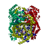
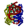
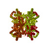
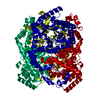
- PDB-7dfk: Crystal structure of xylitol-bound glucose isomerase by serial mi... -
+ Open data

- Basic information
| Entry | Database: PDB / ID: 7dfk | |||||||||
|---|---|---|---|---|---|---|---|---|---|---|
| Title | Crystal structure of xylitol-bound glucose isomerase by serial millisecond crystallography | |||||||||
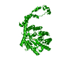
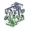
 Components Components | Xylose isomerase | |||||||||
 Keywords Keywords | ISOMERASE / glucose isomerase / xylose isomerase / serial crystallography / serial millisecond crystallography / room temperature / xylitol | |||||||||
| Function / homology |  Function and homology information Function and homology informationxylose isomerase / xylose isomerase activity / D-xylose metabolic process / magnesium ion binding / identical protein binding / cytoplasm Similarity search - Function | |||||||||
| Biological species |  Streptomyces rubiginosus (bacteria) Streptomyces rubiginosus (bacteria) | |||||||||
| Method |  X-RAY DIFFRACTION / SYNCHROTRON / MOLECULAR REPLACEMENT / Resolution: 1.4 Å X-RAY DIFFRACTION / SYNCHROTRON / MOLECULAR REPLACEMENT / Resolution: 1.4 Å | |||||||||
 Authors Authors | Nam, K.H. | |||||||||
| Funding support |  Korea, Republic Of, 2items Korea, Republic Of, 2items
| |||||||||
 Citation Citation | Journal: To Be Published Title: Crystal structure of xylitol-bound glucose isomerase by serial millisecond crystallography Authors: Nam, K.H. | |||||||||
| History |
|
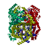
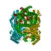
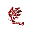
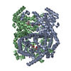
- Structure visualization
| Structure viewer | Molecule: MolmilJmol/JSmol |
|---|
- Downloads & links
-Download
| PDBx/mmCIF format | 7dfk.cif.gz | 161.5 KB | Display | PDBx/mmCIF format |
|---|---|---|---|---|
| PDB format | pdb7dfk.ent.gz | 127.8 KB | Display | PDB format |
| PDBx/mmJSON format | 7dfk.json.gz | Tree view | PDBx/mmJSON format | |
| Others |  Other downloads Other downloads |
-Validation report
| Arichive directory | https://data.pdbj.org/pub/pdb/validation_reports/df/7dfkftp://data.pdbj.org/pub/pdb/validation_reports/df/7dfk | HTTPS FTP |
|---|
-Related structure data
| Related structure data |  7ck0S S: Starting model for refinement |
|---|---|
| Similar structure data | |
| Experimental dataset #1 | Data reference: 10.11577/1777828 / Data set type: diffraction image data |
-Links
 PDBj
PDBj
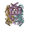
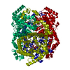
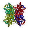
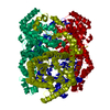
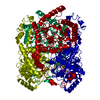
- Assembly
| Deposited unit | 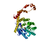
| |||||||||
|---|---|---|---|---|---|---|---|---|---|---|
| 1 | 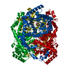
| |||||||||
| Unit cell |
| |||||||||
| Components on special symmetry positions |
|
-Components
| #1: Protein | Mass: 43283.297 Da / Num. of mol.: 1 / Source method: isolated from a natural source / Source: (natural) Streptomyces rubiginosus (bacteria) / References: UniProt: P24300, xylose isomerase |
|---|---|
| #2: Chemical | ChemComp-MG /   Mass: 24.305 Da / Num. of mol.: 1 / Source method: obtained synthetically / Formula: Mg / Feature type: SUBJECT OF INVESTIGATION Mass: 24.305 Da / Num. of mol.: 1 / Source method: obtained synthetically / Formula: Mg / Feature type: SUBJECT OF INVESTIGATION |
| #3: Sugar | ChemComp-XYL /   Type: D-saccharide / Mass: 152.146 Da / Num. of mol.: 1 / Source method: obtained synthetically / Formula: C5H12O5 / Feature type: SUBJECT OF INVESTIGATION Type: D-saccharide / Mass: 152.146 Da / Num. of mol.: 1 / Source method: obtained synthetically / Formula: C5H12O5 / Feature type: SUBJECT OF INVESTIGATION |
| #4: Water | ChemComp-HOH /  Mass: 18.015 Da / Num. of mol.: 314 / Source method: isolated from a natural source / Formula: H2O Mass: 18.015 Da / Num. of mol.: 314 / Source method: isolated from a natural source / Formula: H2O |
| Has ligand of interest | Y |
-Experimental details
-Experiment
| Experiment | Method: X-RAY DIFFRACTION / Number of used crystals: 1 |
|---|
- Sample preparation
| Crystal | Density Matthews: 2.8 Å3/Da / Density % sol: 56.11 % |
|---|---|
| Crystal grow | Temperature: 293.5 K / Method: batch mode / pH: 7 / Details: Tris-HCl, Ammonium sulfate, Magnesium sulfate |
-Data collection
| Diffraction | Mean temperature: 298.15 K / Serial crystal experiment: Y |
|---|---|
| Diffraction source | Source: SYNCHROTRON / Site: PAL/PLS / Beamline: 11C / Wavelength: 0.97942 Å |
| Detector | Type: DECTRIS PILATUS 6M / Detector: PIXEL / Date: Nov 24, 2019 |
| Radiation | Protocol: SINGLE WAVELENGTH / Monochromatic (M) / Laue (L): M / Scattering type: x-ray |
| Radiation wavelength | Wavelength: 0.97942 Å / Relative weight: 1 |
| Reflection | Resolution: 1.4→72.4 Å / Num. obs: 95564 / % possible obs: 100 % / Redundancy: 277.9 % / CC1/2: 0.952 / Net I/σ(I): 4.15 |
| Reflection shell | Resolution: 1.4→1.45 Å / Num. unique obs: 9462 / CC1/2: 0.5968 |
| Serial crystallography sample delivery | Method: fixed target |
| Serial crystallography sample delivery fixed target | Sample holding: nylon mesh / Support base: goniometer |
- Processing
| Software |
| ||||||||||||||||||||||||||||||||||||||||||||||||||||||||||||||||||||||
|---|---|---|---|---|---|---|---|---|---|---|---|---|---|---|---|---|---|---|---|---|---|---|---|---|---|---|---|---|---|---|---|---|---|---|---|---|---|---|---|---|---|---|---|---|---|---|---|---|---|---|---|---|---|---|---|---|---|---|---|---|---|---|---|---|---|---|---|---|---|---|---|
| Refinement | Method to determine structure: MOLECULAR REPLACEMENT Starting model: 7CK0 Resolution: 1.4→71.78 Å / SU ML: 0.23 / Cross valid method: THROUGHOUT / σ(F): 1.33 / Phase error: 29.92 / Stereochemistry target values: ML
| ||||||||||||||||||||||||||||||||||||||||||||||||||||||||||||||||||||||
| Solvent computation | Shrinkage radii: 0.9 Å / VDW probe radii: 1.11 Å / Solvent model: FLAT BULK SOLVENT MODEL | ||||||||||||||||||||||||||||||||||||||||||||||||||||||||||||||||||||||
| Displacement parameters | Biso max: 65.24 Å2 / Biso mean: 21.0188 Å2 / Biso min: 3.82 Å2 | ||||||||||||||||||||||||||||||||||||||||||||||||||||||||||||||||||||||
| Refinement step | Cycle: final / Resolution: 1.4→71.78 Å
| ||||||||||||||||||||||||||||||||||||||||||||||||||||||||||||||||||||||
| LS refinement shell | Refine-ID: X-RAY DIFFRACTION / Rfactor Rfree error: 0 / Total num. of bins used: 9
|
