 Movie
Movie Controller

[English] 日本語
 Yorodumi
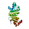
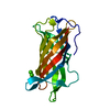
- PDB-7df2: Crystal structure of a C2 domain protein from Ramazzottius varieo... -
+ Open data

- Basic information
| Entry | Database: PDB / ID: 7df2 | |||||||||
|---|---|---|---|---|---|---|---|---|---|---|
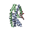
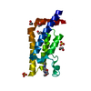
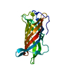
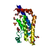
| Title | Crystal structure of a C2 domain protein from Ramazzottius varieornatus | |||||||||
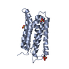
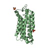
 Components Components | C2 domain protein | |||||||||
 Keywords Keywords | METAL BINDING PROTEIN / tardigrade / Ca-binding protein / C2 domain | |||||||||
| Function / homology | metal ion binding / Uncharacterized protein Function and homology information Function and homology information | |||||||||
| Biological species |  | |||||||||
| Method |  X-RAY DIFFRACTION / SYNCHROTRON / SAD / Resolution: 1.7 Å X-RAY DIFFRACTION / SYNCHROTRON / SAD / Resolution: 1.7 Å | |||||||||
 Authors Authors | Fukuda, Y. / Inoue, T. | |||||||||
| Funding support |  Japan, 2items Japan, 2items
| |||||||||
 Citation Citation | Journal: Protein Sci. / Year: 2021 Title: Structural insights into a C2 domain protein specifically found in tardigrades. Authors: Fukuda, Y. / Inoue, T. | |||||||||
| History |
|
- Structure visualization
| Structure viewer | Molecule: MolmilJmol/JSmol |
|---|
- Downloads & links
-Download
| PDBx/mmCIF format | 7df2.cif.gz | 69.4 KB | Display | PDBx/mmCIF format |
|---|---|---|---|---|
| PDB format | pdb7df2.ent.gz | 39.7 KB | Display | PDB format |
| PDBx/mmJSON format | 7df2.json.gz | Tree view | PDBx/mmJSON format | |
| Others |  Other downloads Other downloads |
-Validation report
| Arichive directory | https://data.pdbj.org/pub/pdb/validation_reports/df/7df2ftp://data.pdbj.org/pub/pdb/validation_reports/df/7df2 | HTTPS FTP |
|---|
-Related structure data
| Similar structure data |
|---|


-Links
 PDBj
PDBj- Assembly
| Deposited unit | 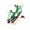
| ||||||||||||
|---|---|---|---|---|---|---|---|---|---|---|---|---|---|
| 1 |
| ||||||||||||
| Unit cell |
| ||||||||||||
| Components on special symmetry positions |
|
-Components
| #1: Protein | Mass: 23879.693 Da / Num. of mol.: 1 Source method: isolated from a genetically manipulated source Source: (gene. exp.) Gene: RvY_00338-1, RvY_00338.1, RvY_00338 / Production host:  | ||||||
|---|---|---|---|---|---|---|---|
| #2: Polysaccharide | beta-D-glucopyranose-(1-1)-alpha-D-glucopyranose Source method: isolated from a genetically manipulated source | ||||||
| #3: Chemical |   Mass: 40.078 Da / Num. of mol.: 3 / Source method: obtained synthetically / Formula: Ca / Feature type: SUBJECT OF INVESTIGATION Mass: 40.078 Da / Num. of mol.: 3 / Source method: obtained synthetically / Formula: Ca / Feature type: SUBJECT OF INVESTIGATION#4: Water | ChemComp-HOH / |  Mass: 18.015 Da / Num. of mol.: 199 / Source method: isolated from a natural source / Formula: H2O Mass: 18.015 Da / Num. of mol.: 199 / Source method: isolated from a natural source / Formula: H2OHas ligand of interest | Y | Has protein modification | Y | |
-Experimental details
-Experiment
| Experiment | Method: X-RAY DIFFRACTION / Number of used crystals: 1 |
|---|
- Sample preparation
| Crystal | Density Matthews: 2.08 Å3/Da / Density % sol: 40.93 % |
|---|---|
| Crystal grow | Temperature: 293 K / Method: vapor diffusion, hanging drop / pH: 6.5 Details: 30 mg per mL protein, 0.1 M calcium chloride dihydrate, 0.1 M Tris pH 6.5, 13% (w/v) polyethylene glycol monomethyl ether 2000 |
-Data collection
| Diffraction | Mean temperature: 100 K / Serial crystal experiment: N |
|---|---|
| Diffraction source | Source: SYNCHROTRON / Site: SPring-8 / Beamline: BL44XU / Wavelength: 0.9 Å |
| Detector | Type: DECTRIS EIGER X 16M / Detector: PIXEL / Date: Nov 3, 2020 |
| Radiation | Protocol: SINGLE WAVELENGTH / Monochromatic (M) / Laue (L): M / Scattering type: x-ray |
| Radiation wavelength | Wavelength: 0.9 Å / Relative weight: 1 |
| Reflection | Resolution: 1.7→40.7 Å / Num. obs: 22431 / % possible obs: 98.4 % / Redundancy: 13.3 % / Biso Wilson estimate: 24.29 Å2 / CC1/2: 0.999 / Rrim(I) all: 0.091 / Net I/σ(I): 16.2 |
| Reflection shell | Resolution: 1.7→1.73 Å / Mean I/σ(I) obs: 2.5 / Num. unique obs: 1128 / CC1/2: 0.87 / Rrim(I) all: 1.29 |
- Processing
| Software |
| |||||||||||||||||||||||||||||||||||||||||||||||||||||||||||||||
|---|---|---|---|---|---|---|---|---|---|---|---|---|---|---|---|---|---|---|---|---|---|---|---|---|---|---|---|---|---|---|---|---|---|---|---|---|---|---|---|---|---|---|---|---|---|---|---|---|---|---|---|---|---|---|---|---|---|---|---|---|---|---|---|---|
| Refinement | Method to determine structure: SAD / Resolution: 1.7→40.68 Å / SU ML: 0.1663 / Cross valid method: FREE R-VALUE / σ(F): 1.34 / Phase error: 21.5273 Stereochemistry target values: GeoStd + Monomer Library + CDL v1.2
| |||||||||||||||||||||||||||||||||||||||||||||||||||||||||||||||
| Solvent computation | Shrinkage radii: 0.9 Å / VDW probe radii: 1.11 Å / Solvent model: FLAT BULK SOLVENT MODEL | |||||||||||||||||||||||||||||||||||||||||||||||||||||||||||||||
| Displacement parameters | Biso mean: 29.35 Å2 | |||||||||||||||||||||||||||||||||||||||||||||||||||||||||||||||
| Refinement step | Cycle: LAST / Resolution: 1.7→40.68 Å
| |||||||||||||||||||||||||||||||||||||||||||||||||||||||||||||||
| Refine LS restraints |
| |||||||||||||||||||||||||||||||||||||||||||||||||||||||||||||||
| LS refinement shell |
|
