 Movie
Movie Controller

[English] 日本語
 Yorodumi
- PDB-7dex: Crystal Structures of Anthocyanin 5,3'-aromatic acyltransferase H... -
+ Open data

- Basic information
| Entry | Database: PDB / ID: 7dex | ||||||
|---|---|---|---|---|---|---|---|
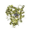
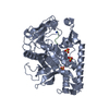
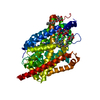
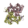
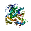
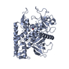
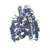
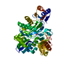
| Title | Crystal Structures of Anthocyanin 5,3'-aromatic acyltransferase H174A mutant with caffeoyl-CoA | ||||||
 Components Components | Anthocyanin 5-aromatic acyltransferase | ||||||
 Keywords Keywords | TRANSFERASE / acyltransferase / anthocyanin / plant enzyme / delphinidin / caffeoyl-CoA | ||||||
| Function / homology | anthocyanin 5-(6'''-hydroxycinnamoyltransferase) / anthocyanin 5-(6'''-hydroxycinnamoyltransferase) activity / : / Transferase family / Chloramphenicol acetyltransferase-like domain superfamily / cytoplasm / Chem-H5L / Anthocyanin 5-aromatic acyltransferase Function and homology information Function and homology information | ||||||
| Biological species |  Gentiana triflora (plant) Gentiana triflora (plant) | ||||||
| Method |  X-RAY DIFFRACTION / SYNCHROTRON / MOLECULAR REPLACEMENT / Resolution: 2.5 Å X-RAY DIFFRACTION / SYNCHROTRON / MOLECULAR REPLACEMENT / Resolution: 2.5 Å | ||||||
 Authors Authors | Murayama, K. / Kato-Murayama, M. / Shirouzu, M. | ||||||
 Citation Citation | Journal: Phytochemistry / Year: 2021 Title: Anthocyanin 5,3'-aromatic acyltransferase from Gentiana triflora, a structural insight into biosynthesis of a blue anthocyanin. Authors: Murayama, K. / Kato-Murayama, M. / Sato, T. / Hosaka, T. / Ishiguro, K. / Mizuno, T. / Kitao, K. / Honma, T. / Yokoyama, S. / Tanaka, Y. / Shirouzu, M. | ||||||
| History |
|
- Structure visualization
| Structure viewer | Molecule: MolmilJmol/JSmol |
|---|
- Downloads & links
-Download
| PDBx/mmCIF format | 7dex.cif.gz | 111.1 KB | Display | PDBx/mmCIF format |
|---|---|---|---|---|
| PDB format | pdb7dex.ent.gz | 81 KB | Display | PDB format |
| PDBx/mmJSON format | 7dex.json.gz | Tree view | PDBx/mmJSON format | |
| Others |  Other downloads Other downloads |
-Validation report
| Summary document | 7dex_validation.pdf.gz | 624.8 KB | Display | wwPDB validaton report |
|---|---|---|---|---|
| Full document | 7dex_full_validation.pdf.gz | 637.5 KB | Display | |
| Data in XML | 7dex_validation.xml.gz | 20.6 KB | Display | |
| Data in CIF | 7dex_validation.cif.gz | 28 KB | Display | |
| Arichive directory | https://data.pdbj.org/pub/pdb/validation_reports/de/7dexftp://data.pdbj.org/pub/pdb/validation_reports/de/7dex | HTTPS FTP |
-Related structure data
| Related structure data | 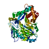 7devC  2e1uS S: Starting model for refinement C: citing same article ( |
|---|---|
| Similar structure data |


-Links
 PDBj
PDBj

- Assembly
| Deposited unit | 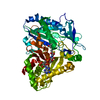
| ||||||||||||
|---|---|---|---|---|---|---|---|---|---|---|---|---|---|
| 1 |
| ||||||||||||
| Unit cell |
|
-Components
| #1: Protein | Mass: 52339.984 Da / Num. of mol.: 1 / Mutation: H174A Source method: isolated from a genetically manipulated source Source: (gene. exp.) Gentiana triflora (plant) / Production host:  References: UniProt: Q9ZWR8, anthocyanin 5-(6'''-hydroxycinnamoyltransferase) |
|---|---|
| #2: Chemical | ChemComp-H5L /   Mass: 929.676 Da / Num. of mol.: 1 / Source method: obtained synthetically / Formula: C30H42N7O19P3S / Feature type: SUBJECT OF INVESTIGATION Mass: 929.676 Da / Num. of mol.: 1 / Source method: obtained synthetically / Formula: C30H42N7O19P3S / Feature type: SUBJECT OF INVESTIGATION |
| #3: Water | ChemComp-HOH /  Mass: 18.015 Da / Num. of mol.: 71 / Source method: isolated from a natural source / Formula: H2O Mass: 18.015 Da / Num. of mol.: 71 / Source method: isolated from a natural source / Formula: H2O |
| Has ligand of interest | Y |
-Experimental details
-Experiment
| Experiment | Method: X-RAY DIFFRACTION / Number of used crystals: 1 |
|---|
- Sample preparation
| Crystal | Density Matthews: 2.72 Å3/Da / Density % sol: 54.78 % |
|---|---|
| Crystal grow | Temperature: 293 K / Method: vapor diffusion, sitting drop / Details: Hepes, sodium citrate |
-Data collection
| Diffraction | Mean temperature: 100 K / Serial crystal experiment: N |
|---|---|
| Diffraction source | Source: SYNCHROTRON / Site: SLS  / Beamline: X10SA / Wavelength: 0.978 Å / Beamline: X10SA / Wavelength: 0.978 Å |
| Detector | Type: DECTRIS PILATUS 6M / Detector: PIXEL / Date: Aug 10, 2012 |
| Radiation | Protocol: SINGLE WAVELENGTH / Monochromatic (M) / Laue (L): M / Scattering type: x-ray |
| Radiation wavelength | Wavelength: 0.978 Å / Relative weight: 1 |
| Reflection | Resolution: 2.5→29.65 Å / Num. obs: 19939 / % possible obs: 99.8 % / Redundancy: 6.6 % / Biso Wilson estimate: 51.12 Å2 / Rmerge(I) obs: 0.11 / Net I/σ(I): 14.2 |
| Reflection shell | Resolution: 2.5→2.64 Å / Rmerge(I) obs: 1.107 / Num. unique obs: 2826 |
- Processing
| Software |
| ||||||||||||||||||||||||||||||||||||||||||||||||||||||||
|---|---|---|---|---|---|---|---|---|---|---|---|---|---|---|---|---|---|---|---|---|---|---|---|---|---|---|---|---|---|---|---|---|---|---|---|---|---|---|---|---|---|---|---|---|---|---|---|---|---|---|---|---|---|---|---|---|---|
| Refinement | Method to determine structure: MOLECULAR REPLACEMENT Starting model: 2E1U Resolution: 2.5→29.65 Å / SU ML: 0.3628 / Cross valid method: FREE R-VALUE / σ(F): 0 / Phase error: 28.7564 / Stereochemistry target values: GeoStd + Monomer Library
| ||||||||||||||||||||||||||||||||||||||||||||||||||||||||
| Solvent computation | Shrinkage radii: 0.9 Å / VDW probe radii: 1.11 Å / Solvent model: FLAT BULK SOLVENT MODEL | ||||||||||||||||||||||||||||||||||||||||||||||||||||||||
| Displacement parameters | Biso mean: 57.34 Å2 | ||||||||||||||||||||||||||||||||||||||||||||||||||||||||
| Refinement step | Cycle: LAST / Resolution: 2.5→29.65 Å
| ||||||||||||||||||||||||||||||||||||||||||||||||||||||||
| Refine LS restraints |
| ||||||||||||||||||||||||||||||||||||||||||||||||||||||||
| LS refinement shell |
|
