 Movie
Movie Controller

[English] 日本語
 Yorodumi
- PDB-7deo: Crystal structure of SARS-CoV-2 RBD in complex with a neutralizin... -
+ Open data

- Basic information
| Entry | Database: PDB / ID: 7deo | |||||||||
|---|---|---|---|---|---|---|---|---|---|---|
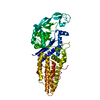
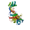
| Title | Crystal structure of SARS-CoV-2 RBD in complex with a neutralizing antibody scFv | |||||||||
 Components Components |
| |||||||||
 Keywords Keywords | VIRAL PROTEIN/ANTIVIRAL PROTEIN / SARS-CoV-2 / RBD / antibody / VIRAL PROTEIN / ANTIVIRAL PROTEIN / VIRAL PROTEIN-ANTIVIRAL PROTEIN complex | |||||||||
| Function / homology |  Function and homology information Function and homology informationsymbiont-mediated disruption of host tissue / Maturation of spike protein / Translation of Structural Proteins / Virion Assembly and Release / host cell surface / host extracellular region / symbiont-mediated-mediated suppression of host tetherin activity / Induction of Cell-Cell Fusion / structural constituent of virion / positive regulation of viral entry into host cell ...symbiont-mediated disruption of host tissue / Maturation of spike protein / Translation of Structural Proteins / Virion Assembly and Release / host cell surface / host extracellular region / symbiont-mediated-mediated suppression of host tetherin activity / Induction of Cell-Cell Fusion / structural constituent of virion / positive regulation of viral entry into host cell / membrane fusion / host cell endoplasmic reticulum-Golgi intermediate compartment membrane / Attachment and Entry / entry receptor-mediated virion attachment to host cell / receptor-mediated virion attachment to host cell / host cell surface receptor binding / symbiont-mediated suppression of host innate immune response / endocytosis involved in viral entry into host cell / receptor ligand activity / fusion of virus membrane with host plasma membrane / fusion of virus membrane with host endosome membrane / viral envelope / symbiont entry into host cell / virion attachment to host cell / host cell plasma membrane / SARS-CoV-2 activates/modulates innate and adaptive immune responses / virion membrane / membrane / identical protein binding / plasma membrane Similarity search - Function | |||||||||
| Biological species |  Homo sapiens (human) Homo sapiens (human)  Severe acute respiratory syndrome coronavirus 2 Severe acute respiratory syndrome coronavirus 2 | |||||||||
| Method |  X-RAY DIFFRACTION / SYNCHROTRON / MOLECULAR REPLACEMENT / Resolution: 2.5 Å X-RAY DIFFRACTION / SYNCHROTRON / MOLECULAR REPLACEMENT / Resolution: 2.5 Å | |||||||||
 Authors Authors | Fu, D. / Zhang, G. / Li, X. / Rao, Z. / Guo, Y. | |||||||||
| Funding support |  China, 2items China, 2items
| |||||||||
 Citation Citation | Journal: Plos Biol. / Year: 2021 Title: Structural basis for SARS-CoV-2 neutralizing antibodies with novel binding epitopes. Authors: Fu, D. / Zhang, G. / Wang, Y. / Zhang, Z. / Hu, H. / Shen, S. / Wu, J. / Li, B. / Li, X. / Fang, Y. / Liu, J. / Wang, Q. / Zhou, Y. / Wang, W. / Li, Y. / Lu, Z. / Wang, X. / Nie, C. / Tian, ...Authors: Fu, D. / Zhang, G. / Wang, Y. / Zhang, Z. / Hu, H. / Shen, S. / Wu, J. / Li, B. / Li, X. / Fang, Y. / Liu, J. / Wang, Q. / Zhou, Y. / Wang, W. / Li, Y. / Lu, Z. / Wang, X. / Nie, C. / Tian, Y. / Chen, D. / Wang, Y. / Zhou, X. / Wang, Q. / Yu, F. / Zhang, C. / Deng, C. / Zhou, L. / Guan, G. / Shao, N. / Lou, Z. / Deng, F. / Zhang, H. / Chen, X. / Wang, M. / Liu, L. / Rao, Z. / Guo, Y. | |||||||||
| History |
|
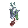
- Structure visualization
| Structure viewer | Molecule: MolmilJmol/JSmol |
|---|
- Downloads & links
-Download
| PDBx/mmCIF format | 7deo.cif.gz | 189.5 KB | Display | PDBx/mmCIF format |
|---|---|---|---|---|
| PDB format | pdb7deo.ent.gz | 147.1 KB | Display | PDB format |
| PDBx/mmJSON format | 7deo.json.gz | Tree view | PDBx/mmJSON format | |
| Others |  Other downloads Other downloads |
-Validation report
| Arichive directory | https://data.pdbj.org/pub/pdb/validation_reports/de/7deoftp://data.pdbj.org/pub/pdb/validation_reports/de/7deo | HTTPS FTP |
|---|
-Related structure data
| Related structure data | 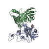 7detC 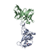 7deuC  7cjfS S: Starting model for refinement C: citing same article ( |
|---|---|
| Similar structure data |


-Links
 PDBj
PDBj


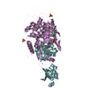
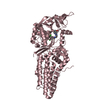
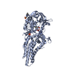
- Assembly
| Deposited unit | 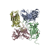
| ||||||||
|---|---|---|---|---|---|---|---|---|---|
| 1 | 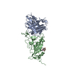
| ||||||||
| 2 | 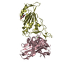
| ||||||||
| Unit cell |
|
-Components
| #1: Antibody | Mass: 27654.650 Da / Num. of mol.: 2 Source method: isolated from a genetically manipulated source Source: (gene. exp.) Homo sapiens (human) / Production host: Homo sapiens (human)#2: Protein | Mass: 23159.947 Da / Num. of mol.: 2 / Fragment: RBD Source method: isolated from a genetically manipulated source Source: (gene. exp.) Severe acute respiratory syndrome coronavirus 2Gene: S, 2 / Production host:   Spodoptera frugiperda (fall armyworm) / References: UniProt: P0DTC2 Spodoptera frugiperda (fall armyworm) / References: UniProt: P0DTC2#3: Chemical |   Mass: 40.078 Da / Num. of mol.: 2 / Source method: obtained synthetically / Formula: Ca Mass: 40.078 Da / Num. of mol.: 2 / Source method: obtained synthetically / Formula: Ca#4: Sugar |   Type: D-saccharide, beta linking / Mass: 221.208 Da / Num. of mol.: 2 / Source method: obtained synthetically / Formula: C8H15NO6 Type: D-saccharide, beta linking / Mass: 221.208 Da / Num. of mol.: 2 / Source method: obtained synthetically / Formula: C8H15NO6#5: Water | ChemComp-HOH / |  Mass: 18.015 Da / Num. of mol.: 325 / Source method: isolated from a natural source / Formula: H2O Mass: 18.015 Da / Num. of mol.: 325 / Source method: isolated from a natural source / Formula: H2OHas ligand of interest | N | Has protein modification | Y | |
|---|
-Experimental details
-Experiment
| Experiment | Method: X-RAY DIFFRACTION / Number of used crystals: 1 |
|---|
- Sample preparation
| Crystal | Density Matthews: 2.63 Å3/Da / Density % sol: 53.2 % |
|---|---|
| Crystal grow | Temperature: 293 K / Method: vapor diffusion Details: 1% w/v Tryptone, 0.001M Sodium azide, 0.05M HEPES sodium pH 7.0, 12% w/v Polyethylene glycol 3350 |
-Data collection
| Diffraction | Mean temperature: 100 K / Serial crystal experiment: N |
|---|---|
| Diffraction source | Source: SYNCHROTRON / Site: SwissFEL ARAMIS  / Beamline: ESA / Wavelength: 1 Å / Beamline: ESA / Wavelength: 1 Å |
| Detector | Type: DECTRIS EIGER X 16M / Detector: PIXEL / Date: Sep 25, 2020 |
| Radiation | Protocol: SINGLE WAVELENGTH / Monochromatic (M) / Laue (L): M / Scattering type: x-ray |
| Radiation wavelength | Wavelength: 1 Å / Relative weight: 1 |
| Reflection | Resolution: 2.5→50 Å / Num. obs: 70511 / % possible obs: 99.4 % / Redundancy: 4.4 % / CC1/2: 0.979 / Rmerge(I) obs: 0.128 / Net I/σ(I): 7.2 |
| Reflection shell | Resolution: 2.5→2.6 Å / Rmerge(I) obs: 0.525 / Num. unique obs: 4161 / CC1/2: 0.639 |
- Processing
| Software |
| |||||||||||||||||||||||||||||||||||||||||||||||||||||||||||||||||||||||||||||||||||||||||||||||||||||||||||||||||||||||||||||||||||||||||||||||||||||||||||||||||||||||||||||||||||||||||||||
|---|---|---|---|---|---|---|---|---|---|---|---|---|---|---|---|---|---|---|---|---|---|---|---|---|---|---|---|---|---|---|---|---|---|---|---|---|---|---|---|---|---|---|---|---|---|---|---|---|---|---|---|---|---|---|---|---|---|---|---|---|---|---|---|---|---|---|---|---|---|---|---|---|---|---|---|---|---|---|---|---|---|---|---|---|---|---|---|---|---|---|---|---|---|---|---|---|---|---|---|---|---|---|---|---|---|---|---|---|---|---|---|---|---|---|---|---|---|---|---|---|---|---|---|---|---|---|---|---|---|---|---|---|---|---|---|---|---|---|---|---|---|---|---|---|---|---|---|---|---|---|---|---|---|---|---|---|---|---|---|---|---|---|---|---|---|---|---|---|---|---|---|---|---|---|---|---|---|---|---|---|---|---|---|---|---|---|---|---|---|---|
| Refinement | Method to determine structure: MOLECULAR REPLACEMENT Starting model: 7CJF Resolution: 2.5→49.49 Å / SU ML: 0.36 / Cross valid method: THROUGHOUT / σ(F): 1.34 / Phase error: 24.41 / Stereochemistry target values: ML
| |||||||||||||||||||||||||||||||||||||||||||||||||||||||||||||||||||||||||||||||||||||||||||||||||||||||||||||||||||||||||||||||||||||||||||||||||||||||||||||||||||||||||||||||||||||||||||||
| Solvent computation | Shrinkage radii: 0.9 Å / VDW probe radii: 1.11 Å / Solvent model: FLAT BULK SOLVENT MODEL | |||||||||||||||||||||||||||||||||||||||||||||||||||||||||||||||||||||||||||||||||||||||||||||||||||||||||||||||||||||||||||||||||||||||||||||||||||||||||||||||||||||||||||||||||||||||||||||
| Displacement parameters | Biso max: 102.96 Å2 / Biso mean: 32.439 Å2 / Biso min: 12.37 Å2 | |||||||||||||||||||||||||||||||||||||||||||||||||||||||||||||||||||||||||||||||||||||||||||||||||||||||||||||||||||||||||||||||||||||||||||||||||||||||||||||||||||||||||||||||||||||||||||||
| Refinement step | Cycle: final / Resolution: 2.5→49.49 Å
| |||||||||||||||||||||||||||||||||||||||||||||||||||||||||||||||||||||||||||||||||||||||||||||||||||||||||||||||||||||||||||||||||||||||||||||||||||||||||||||||||||||||||||||||||||||||||||||
| LS refinement shell | Refine-ID: X-RAY DIFFRACTION / Rfactor Rfree error: 0 / Total num. of bins used: 26
|
