 Movie
Movie Controller

[English] 日本語
 Yorodumi
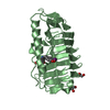
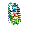
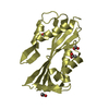
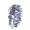
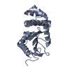
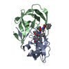
- PDB-7ddb: Crystal structure of fungal antifreeze protein with intermediate ... -
+ Open data

- Basic information
| Entry | Database: PDB / ID: 7ddb | ||||||
|---|---|---|---|---|---|---|---|
| Title | Crystal structure of fungal antifreeze protein with intermediate activity | ||||||
 Components Components | Antifreeze protein | ||||||
 Keywords Keywords | ANTIFREEZE PROTEIN / right-handed beta-helix / beta-solenoid / ice-binding protein / thermal hysteresis protein / ice-structuring protein | ||||||
| Function / homology | Ice-binding protein / Ice-binding-like / Antifreeze protein Function and homology information Function and homology information | ||||||
| Biological species |  Typhula ishikariensis (fungus) Typhula ishikariensis (fungus) | ||||||
| Method |  X-RAY DIFFRACTION / SYNCHROTRON / MOLECULAR REPLACEMENT / molecular replacement / Resolution: 1.72 Å X-RAY DIFFRACTION / SYNCHROTRON / MOLECULAR REPLACEMENT / molecular replacement / Resolution: 1.72 Å | ||||||
 Authors Authors | Khan, N.M.M.U. / Arai, T. / Tsuda, S. / Kondo, H. | ||||||
| Funding support |  Japan, 1items Japan, 1items
| ||||||
 Citation Citation | Journal: Sci Rep / Year: 2021 Title: Characterization of microbial antifreeze protein with intermediate activity suggests that a bound-water network is essential for hyperactivity. Authors: Khan, N.M.U. / Arai, T. / Tsuda, S. / Kondo, H. | ||||||
| History |
|
- Structure visualization
| Structure viewer | Molecule: MolmilJmol/JSmol |
|---|
- Downloads & links
-Download
| PDBx/mmCIF format | 7ddb.cif.gz | 104.6 KB | Display | PDBx/mmCIF format |
|---|---|---|---|---|
| PDB format | pdb7ddb.ent.gz | 77 KB | Display | PDB format |
| PDBx/mmJSON format | 7ddb.json.gz | Tree view | PDBx/mmJSON format | |
| Others |  Other downloads Other downloads |
-Validation report
| Arichive directory | https://data.pdbj.org/pub/pdb/validation_reports/dd/7ddbftp://data.pdbj.org/pub/pdb/validation_reports/dd/7ddb | HTTPS FTP |
|---|
-Related structure data
| Related structure data | 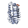 7dc5C  5b5hS C: citing same article ( S: Starting model for refinement |
|---|---|
| Similar structure data |


-Links
 PDBj
PDBj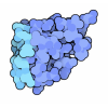
- Assembly
| Deposited unit | 
| ||||||||
|---|---|---|---|---|---|---|---|---|---|
| 1 | 
| ||||||||
| 2 | 
| ||||||||
| Unit cell |
|
-Components
| #1: Protein | Mass: 22089.811 Da / Num. of mol.: 2 / Mutation: T20Y Source method: isolated from a genetically manipulated source Source: (gene. exp.) Typhula ishikariensis (fungus) / Gene: K1-C / Production host:  #2: Chemical |   Mass: 24.305 Da / Num. of mol.: 3 / Source method: obtained synthetically / Formula: Mg Mass: 24.305 Da / Num. of mol.: 3 / Source method: obtained synthetically / Formula: Mg#3: Water | ChemComp-HOH / |  Mass: 18.015 Da / Num. of mol.: 554 / Source method: isolated from a natural source / Formula: H2O Mass: 18.015 Da / Num. of mol.: 554 / Source method: isolated from a natural source / Formula: H2OHas ligand of interest | N | |
|---|
-Experimental details
-Experiment
| Experiment | Method: X-RAY DIFFRACTION / Number of used crystals: 1 |
|---|
- Sample preparation
| Crystal | Density Matthews: 2.07 Å3/Da / Density % sol: 40.65 % / Description: thin plate |
|---|---|
| Crystal grow | Temperature: 293 K / Method: vapor diffusion, hanging drop / pH: 8.5 Details: 0.1M Tris-HCl pH8.5, 30% (w/v) PEG 4000, 0.2M magnesium chloride PH range: 8.5 |
-Data collection
| Diffraction | Mean temperature: 95 K / Serial crystal experiment: N | ||||||||||||||||||||||||||||||
|---|---|---|---|---|---|---|---|---|---|---|---|---|---|---|---|---|---|---|---|---|---|---|---|---|---|---|---|---|---|---|---|
| Diffraction source | Source: SYNCHROTRON / Site: Photon Factory / Beamline: BL-1A / Wavelength: 1.1 Å | ||||||||||||||||||||||||||||||
| Detector | Type: DECTRIS EIGER X 4M / Detector: PIXEL / Date: Mar 17, 2019 | ||||||||||||||||||||||||||||||
| Radiation | Protocol: SINGLE WAVELENGTH / Monochromatic (M) / Laue (L): M / Scattering type: x-ray | ||||||||||||||||||||||||||||||
| Radiation wavelength | Wavelength: 1.1 Å / Relative weight: 1 | ||||||||||||||||||||||||||||||
| Reflection | Resolution: 1.7→49.68 Å / Num. obs: 41109 / % possible obs: 99.8 % / Redundancy: 12.9 % / CC1/2: 0.98 / Rmerge(I) obs: 0.192 / Rpim(I) all: 0.057 / Rrim(I) all: 0.201 / Net I/σ(I): 8.6 | ||||||||||||||||||||||||||||||
| Reflection shell | Diffraction-ID: 1
|
-Phasing
| Phasing | Method: molecular replacement | |||||||||
|---|---|---|---|---|---|---|---|---|---|---|
| Phasing MR |
|
- Processing
| Software |
| ||||||||||||||||||||||||||||||||||||||||||||||||||||||||||||
|---|---|---|---|---|---|---|---|---|---|---|---|---|---|---|---|---|---|---|---|---|---|---|---|---|---|---|---|---|---|---|---|---|---|---|---|---|---|---|---|---|---|---|---|---|---|---|---|---|---|---|---|---|---|---|---|---|---|---|---|---|---|
| Refinement | Method to determine structure: MOLECULAR REPLACEMENT Starting model: 5B5H Resolution: 1.72→49.68 Å / Cor.coef. Fo:Fc: 0.94 / Cor.coef. Fo:Fc free: 0.92 / SU B: 3.005 / SU ML: 0.099 / SU R Cruickshank DPI: 0.1457 / Cross valid method: THROUGHOUT / σ(F): 0 / ESU R: 0.146 / ESU R Free: 0.134 / Stereochemistry target values: MAXIMUM LIKELIHOOD Details: HYDROGENS HAVE BEEN ADDED IN THE RIDING POSITIONS U VALUES : REFINED INDIVIDUALLY
| ||||||||||||||||||||||||||||||||||||||||||||||||||||||||||||
| Solvent computation | Ion probe radii: 0.8 Å / Shrinkage radii: 0.8 Å / VDW probe radii: 1.2 Å / Solvent model: MASK | ||||||||||||||||||||||||||||||||||||||||||||||||||||||||||||
| Displacement parameters | Biso max: 62.39 Å2 / Biso mean: 10.893 Å2 / Biso min: 2.36 Å2
| ||||||||||||||||||||||||||||||||||||||||||||||||||||||||||||
| Refinement step | Cycle: final / Resolution: 1.72→49.68 Å
| ||||||||||||||||||||||||||||||||||||||||||||||||||||||||||||
| Refine LS restraints |
| ||||||||||||||||||||||||||||||||||||||||||||||||||||||||||||
| LS refinement shell | Resolution: 1.72→1.765 Å / Rfactor Rfree error: 0 / Total num. of bins used: 20
|
