 Movie
Movie Controller


+ Open data

- Basic information
| Entry | Database: PDB / ID: 7dbe | ||||||
|---|---|---|---|---|---|---|---|
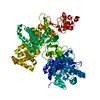
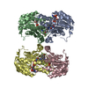
| Title | Structure of a novel transaminase | ||||||
 Components Components | branched-chain amino acid aminotransferase | ||||||
 Keywords Keywords | TRANSFERASE / transaminase | ||||||
| Function / homology | D-amino Acid Aminotransferase; Chain A, domain 2 / D-amino Acid Aminotransferase, subunit A, domain 2 / Alpha-Beta Barrel / Alpha Beta / PYRIDOXAL-5'-PHOSPHATE Function and homology information Function and homology information | ||||||
| Biological species |  Rhodobacter sp. 140A (bacteria) Rhodobacter sp. 140A (bacteria) | ||||||
| Method |  X-RAY DIFFRACTION / SYNCHROTRON / MOLECULAR REPLACEMENT / Resolution: 1.9 Å X-RAY DIFFRACTION / SYNCHROTRON / MOLECULAR REPLACEMENT / Resolution: 1.9 Å | ||||||
 Authors Authors | Li, F.L. / Yu, H.M. | ||||||
 Citation Citation | Journal: Adv.Synth.Catal. / Year: 2021 Title: Biochemical and Structural Characterization of an (R)-Selective Transaminase in the Asymmetric Synthesis of Chiral Hydroxy Amines. Authors: Li, F.L. / Liang, Y.X. / Wei, Y.W. / Zheng, Y.K. / Yu, H.M. | ||||||
| History |
|
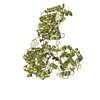
- Structure visualization
| Structure viewer | Molecule: MolmilJmol/JSmol |
|---|
- Downloads & links
-Download
| PDBx/mmCIF format | 7dbe.cif.gz | 366.1 KB | Display | PDBx/mmCIF format |
|---|---|---|---|---|
| PDB format | pdb7dbe.ent.gz | 236.8 KB | Display | PDB format |
| PDBx/mmJSON format | 7dbe.json.gz | Tree view | PDBx/mmJSON format | |
| Others |  Other downloads Other downloads |
-Validation report
| Summary document | 7dbe_validation.pdf.gz | 1.5 MB | Display | wwPDB validaton report |
|---|---|---|---|---|
| Full document | 7dbe_full_validation.pdf.gz | 1.5 MB | Display | |
| Data in XML | 7dbe_validation.xml.gz | 63.3 KB | Display | |
| Data in CIF | 7dbe_validation.cif.gz | 96.1 KB | Display | |
| Arichive directory | https://data.pdbj.org/pub/pdb/validation_reports/db/7dbeftp://data.pdbj.org/pub/pdb/validation_reports/db/7dbe | HTTPS FTP |
-Related structure data
| Related structure data |  3wwhS S: Starting model for refinement |
|---|---|
| Similar structure data |


-Links
 PDBj
PDBj- Assembly
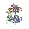
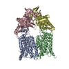
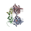
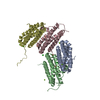
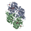
| Deposited unit | 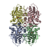
| ||||||||||||
|---|---|---|---|---|---|---|---|---|---|---|---|---|---|
| 1 |
| ||||||||||||
| Unit cell |
|
-Components
| #1: Protein | Mass: 37684.895 Da / Num. of mol.: 4 Source method: isolated from a genetically manipulated source Source: (gene. exp.) Rhodobacter sp. 140A (bacteria) / Production host: #2: Chemical | ChemComp-PLP /   Mass: 247.142 Da / Num. of mol.: 4 / Source method: obtained synthetically / Formula: C8H10NO6P / Feature type: SUBJECT OF INVESTIGATION Mass: 247.142 Da / Num. of mol.: 4 / Source method: obtained synthetically / Formula: C8H10NO6P / Feature type: SUBJECT OF INVESTIGATION#3: Chemical | ChemComp-EDO /   Mass: 62.068 Da / Num. of mol.: 4 / Source method: obtained synthetically / Formula: C2H6O2 Mass: 62.068 Da / Num. of mol.: 4 / Source method: obtained synthetically / Formula: C2H6O2#4: Water | ChemComp-HOH / |  Mass: 18.015 Da / Num. of mol.: 1577 / Source method: isolated from a natural source / Formula: H2O Mass: 18.015 Da / Num. of mol.: 1577 / Source method: isolated from a natural source / Formula: H2OHas ligand of interest | Y | Sequence details | Sequence has been deposited to Genbank with accession ID RBP85366. N-terminal residues MHHHHHH are ...Sequence has been deposited to Genbank with accession ID RBP85366. N-terminal residues MHHHHHH are from expression tag. | |
|---|
-Experimental details
-Experiment
| Experiment | Method: X-RAY DIFFRACTION / Number of used crystals: 1 |
|---|
- Sample preparation
| Crystal | Density Matthews: 2.67 Å3/Da / Density % sol: 53.96 % |
|---|---|
| Crystal grow | Temperature: 291 K / Method: vapor diffusion, sitting drop Details: 2% v/v Tacsimate pH 7.0, 5% v/v 2-Propanol, 0.1M Imidazole pH 7.0, 8% w/v Polyethylene glycol 3350 |
-Data collection
| Diffraction | Mean temperature: 100 K / Serial crystal experiment: N |
|---|---|
| Diffraction source | Source: SYNCHROTRON / Site: SSRF  / Beamline: BL19U1 / Wavelength: 0.9789 Å / Beamline: BL19U1 / Wavelength: 0.9789 Å |
| Detector | Type: DECTRIS PILATUS3 S 6M / Detector: PIXEL / Date: Dec 15, 2019 |
| Radiation | Protocol: SINGLE WAVELENGTH / Monochromatic (M) / Laue (L): M / Scattering type: x-ray |
| Radiation wavelength | Wavelength: 0.9789 Å / Relative weight: 1 |
| Reflection | Resolution: 1.9→50 Å / Num. obs: 120439 / % possible obs: 99.6 % / Redundancy: 6.8 % / Biso Wilson estimate: 22.61 Å2 / CC1/2: 0.962 / Rmerge(I) obs: 0.131 / Rpim(I) all: 0.053 / Rrim(I) all: 0.142 / Rsym value: 0.131 / Net I/σ(I): 18.7 |
| Reflection shell | Resolution: 1.9→1.93 Å / Rmerge(I) obs: 0.665 / Mean I/σ(I) obs: 2.8 / Num. unique obs: 6029 / CC1/2: 0.565 / Rpim(I) all: 0.267 / Rrim(I) all: 0.721 / Rsym value: 0.665 |
- Processing
| Software |
| |||||||||||||||||||||||||||||||||||||||||||||||||||||||||||||||||||||||||||||||||||||||||||||||||||||||||||||||||||||||||||||||||||||||||||||||||||||||||||||||||||||||||||||||||||||||||||||||||||||||||||||||||||||||||
|---|---|---|---|---|---|---|---|---|---|---|---|---|---|---|---|---|---|---|---|---|---|---|---|---|---|---|---|---|---|---|---|---|---|---|---|---|---|---|---|---|---|---|---|---|---|---|---|---|---|---|---|---|---|---|---|---|---|---|---|---|---|---|---|---|---|---|---|---|---|---|---|---|---|---|---|---|---|---|---|---|---|---|---|---|---|---|---|---|---|---|---|---|---|---|---|---|---|---|---|---|---|---|---|---|---|---|---|---|---|---|---|---|---|---|---|---|---|---|---|---|---|---|---|---|---|---|---|---|---|---|---|---|---|---|---|---|---|---|---|---|---|---|---|---|---|---|---|---|---|---|---|---|---|---|---|---|---|---|---|---|---|---|---|---|---|---|---|---|---|---|---|---|---|---|---|---|---|---|---|---|---|---|---|---|---|---|---|---|---|---|---|---|---|---|---|---|---|---|---|---|---|---|---|---|---|---|---|---|---|---|---|---|---|---|---|---|---|---|
| Refinement | Method to determine structure: MOLECULAR REPLACEMENT Starting model: 3WWH Resolution: 1.9→46.85 Å / SU ML: 0.1861 / Cross valid method: FREE R-VALUE / σ(F): 1.35 / Phase error: 19.9076 / Stereochemistry target values: CDL v1.2
| |||||||||||||||||||||||||||||||||||||||||||||||||||||||||||||||||||||||||||||||||||||||||||||||||||||||||||||||||||||||||||||||||||||||||||||||||||||||||||||||||||||||||||||||||||||||||||||||||||||||||||||||||||||||||
| Solvent computation | Shrinkage radii: 0.9 Å / VDW probe radii: 1.11 Å / Solvent model: FLAT BULK SOLVENT MODEL | |||||||||||||||||||||||||||||||||||||||||||||||||||||||||||||||||||||||||||||||||||||||||||||||||||||||||||||||||||||||||||||||||||||||||||||||||||||||||||||||||||||||||||||||||||||||||||||||||||||||||||||||||||||||||
| Displacement parameters | Biso mean: 26.39 Å2 | |||||||||||||||||||||||||||||||||||||||||||||||||||||||||||||||||||||||||||||||||||||||||||||||||||||||||||||||||||||||||||||||||||||||||||||||||||||||||||||||||||||||||||||||||||||||||||||||||||||||||||||||||||||||||
| Refinement step | Cycle: LAST / Resolution: 1.9→46.85 Å
| |||||||||||||||||||||||||||||||||||||||||||||||||||||||||||||||||||||||||||||||||||||||||||||||||||||||||||||||||||||||||||||||||||||||||||||||||||||||||||||||||||||||||||||||||||||||||||||||||||||||||||||||||||||||||
| Refine LS restraints |
| |||||||||||||||||||||||||||||||||||||||||||||||||||||||||||||||||||||||||||||||||||||||||||||||||||||||||||||||||||||||||||||||||||||||||||||||||||||||||||||||||||||||||||||||||||||||||||||||||||||||||||||||||||||||||
| LS refinement shell |
|
