- PDB-7d8v: Crystal Structure of A Kinesin-3 KIF13B mutant-T192Y -
+
Open data
ID or keywords:
Loading...
-
Basic information
Entry
Database: PDB / ID: 7d8v
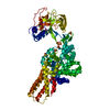
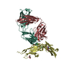
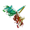
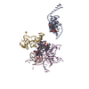
Title
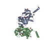
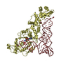
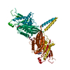
Crystal Structure of A Kinesin-3 KIF13B mutant-T192Y
Components
Kinesin family member 13B
Keywords
TRANSPORT PROTEIN / Kinesin / ATPase
Function / homology
Function and homology information
Kinesins / COPI-dependent Golgi-to-ER retrograde traffic / MHC class II antigen presentation / paranode region of axon / vesicle transport along microtubule / microtubule motor activity / kinesin complex / microtubule-based movement / regulation of axonogenesis / microvillus ...Kinesins / COPI-dependent Golgi-to-ER retrograde traffic / MHC class II antigen presentation / paranode region of axon / vesicle transport along microtubule / microtubule motor activity / kinesin complex / microtubule-based movement / regulation of axonogenesis / microvillus / 14-3-3 protein binding / establishment of protein localization / microtubule binding / microtubule / axon / protein kinase binding / ATP hydrolysis activity / ATP binding / metal ion binding / cytoplasm / cytosol Similarity search - Function
Kinesin-like KIF1-type / Kinesin protein 1B / Kinesin-like / Kinesin protein / Kinesin-associated / Kinesin-associated / CAP-Gly domain signature. / CAP Gly-rich domain / CAP Gly-rich domain superfamily / CAP-Gly domain ...Kinesin-like KIF1-type / Kinesin protein 1B / Kinesin-like / Kinesin protein / Kinesin-associated / Kinesin-associated / CAP-Gly domain signature. / CAP Gly-rich domain / CAP Gly-rich domain superfamily / CAP-Gly domain / CAP-Gly domain profile. / CAP_GLY / Kinesin motor domain / Kinesin / FHA domain / Forkhead-associated (FHA) domain / Kinesin motor domain signature. / Kinesin motor domain, conserved site / Kinesin motor domain / Kinesin motor domain profile. / Kinesin motor, catalytic domain. ATPase. / Kinesin motor domain / SMAD/FHA domain superfamily / Kinesin motor domain superfamily / P-loop containing nucleoside triphosphate hydrolase / 3-Layer(aba) Sandwich / Alpha Beta Similarity search - Domain/homology
In the structure databanks used in Yorodumi, some data are registered as the other names, "COVID-19 virus" and "2019-nCoV". Here are the details of the virus and the list of structure data.
Jan 31, 2019. EMDB accession codes are about to change! (news from PDBe EMDB page)
EMDB accession codes are about to change! (news from PDBe EMDB page)
The allocation of 4 digits for EMDB accession codes will soon come to an end. Whilst these codes will remain in use, new EMDB accession codes will include an additional digit and will expand incrementally as the available range of codes is exhausted. The current 4-digit format prefixed with “EMD-” (i.e. EMD-XXXX) will advance to a 5-digit format (i.e. EMD-XXXXX), and so on. It is currently estimated that the 4-digit codes will be depleted around Spring 2019, at which point the 5-digit format will come into force.
The EM Navigator/Yorodumi systems omit the EMD- prefix.
Related info.:Q: What is EMD? / ID/Accession-code notation in Yorodumi/EM Navigator
Yorodumi is a browser for structure data from EMDB, PDB, SASBDB, etc.
This page is also the successor to EM Navigator detail page, and also detail information page/front-end page for Omokage search.
The word "yorodu" (or yorozu) is an old Japanese word meaning "ten thousand". "mi" (miru) is to see.
Related info.:EMDB / PDB / SASBDB / Comparison of 3 databanks / Yorodumi Search / Aug 31, 2016. New EM Navigator & Yorodumi / Yorodumi Papers / Jmol/JSmol / Function and homology information / Changes in new EM Navigator and Yorodumi
 Movie
Movie Controller


 Open data
 Basic information
 Components
 Keywords
 Function and homology information
 X-RAY DIFFRACTION /
  Authors
 China, 1items
  Citation
 Structure visualization
 Downloads & links
 Other downloads


 PDBj
PDBj


 Assembly
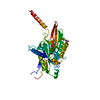


 Mass: 122.143 Da / Num. of mol.: 1 / Source method: obtained synthetically / Formula: C4H12NO3 / Comment: pH buffer*YM
 Mass: 427.201 Da / Num. of mol.: 1 / Source method: obtained synthetically / Formula: C10H15N5O10P2 / Comment: ADP, energy-carrying molecule*YM
 Mass: 24.305 Da / Num. of mol.: 1 / Source method: obtained synthetically / Formula: Mg
 Mass: 18.015 Da / Num. of mol.: 113 / Source method: isolated from a natural source / Formula: H2O
 Sample preparation
 Processing