 Movie
Movie Controller

[English] 日本語
 Yorodumi
- PDB-7d8u: Crystal structure of the C-terminal domain of pNP868R from Africa... -
+ Open data

- Basic information
| Entry | Database: PDB / ID: 7d8u | ||||||
|---|---|---|---|---|---|---|---|
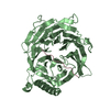
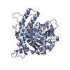
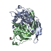
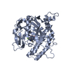
| Title | Crystal structure of the C-terminal domain of pNP868R from African swine fever virus | ||||||
 Components Components | GTP--RNA guanylyltransferase | ||||||
 Keywords Keywords | TRANSFERASE / ASFV / RNA capping / methyltransferase | ||||||
| Function / homology |  Function and homology information Function and homology informationmRNA 5'-triphosphate monophosphatase activity / mRNA 5'-phosphatase / polynucleotide 5'-phosphatase activity / virion component / mRNA guanylyltransferase activity / mRNA guanylyltransferase / mRNA (guanine-N7)-methyltransferase / mRNA 5'-cap (guanine-N7-)-methyltransferase activity / GTP binding / RNA binding Similarity search - Function | ||||||
| Biological species |   African swine fever virus African swine fever virus | ||||||
| Method |  X-RAY DIFFRACTION / SYNCHROTRON / MOLECULAR REPLACEMENT / Resolution: 2.7 Å X-RAY DIFFRACTION / SYNCHROTRON / MOLECULAR REPLACEMENT / Resolution: 2.7 Å | ||||||
 Authors Authors | Du, X. / Geng, Z. / Zhang, H. | ||||||
| Funding support |  China, 1items China, 1items
| ||||||
 Citation Citation | Journal: J.Virol. / Year: 2020 Title: Structure and Biochemical Characteristic of the Methyltransferase (MTase) Domain of RNA Capping Enzyme from African Swine Fever Virus. Authors: Du, X. / Gao, Z.Q. / Geng, Z. / Dong, Y.H. / Zhang, H. | ||||||
| History |
|
- Structure visualization
| Structure viewer | Molecule: MolmilJmol/JSmol |
|---|
- Downloads & links
-Download
| PDBx/mmCIF format | 7d8u.cif.gz | 131.5 KB | Display | PDBx/mmCIF format |
|---|---|---|---|---|
| PDB format | pdb7d8u.ent.gz | 101.1 KB | Display | PDB format |
| PDBx/mmJSON format | 7d8u.json.gz | Tree view | PDBx/mmJSON format | |
| Others |  Other downloads Other downloads |
-Validation report
| Summary document | 7d8u_validation.pdf.gz | 3.1 MB | Display | wwPDB validaton report |
|---|---|---|---|---|
| Full document | 7d8u_full_validation.pdf.gz | 3.1 MB | Display | |
| Data in XML | 7d8u_validation.xml.gz | 23.4 KB | Display | |
| Data in CIF | 7d8u_validation.cif.gz | 31.3 KB | Display | |
| Arichive directory | https://data.pdbj.org/pub/pdb/validation_reports/d8/7d8uftp://data.pdbj.org/pub/pdb/validation_reports/d8/7d8u | HTTPS FTP |
-Related structure data
| Related structure data | 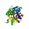 1ri1S S: Starting model for refinement |
|---|---|
| Similar structure data |


-Links
 PDBj
PDBj


- Assembly
| Deposited unit | 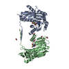
| ||||||||
|---|---|---|---|---|---|---|---|---|---|
| 1 | 
| ||||||||
| 2 | 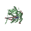
| ||||||||
| Unit cell |
|
-Components
| #1: Protein | Mass: 33056.953 Da / Num. of mol.: 2 Source method: isolated from a genetically manipulated source Source: (gene. exp.) African swine fever virus / Gene: NP868R / Production host:  References: UniProt: A0A0A1DYG7, UniProt: P32094*PLUS, mRNA (guanine-N7)-methyltransferase, mRNA guanylyltransferase, polynucleotide 5'-phosphatase #2: Chemical |   Mass: 398.437 Da / Num. of mol.: 2 / Source method: obtained synthetically / Formula: C15H22N6O5S / Feature type: SUBJECT OF INVESTIGATION Mass: 398.437 Da / Num. of mol.: 2 / Source method: obtained synthetically / Formula: C15H22N6O5S / Feature type: SUBJECT OF INVESTIGATION#3: Chemical | ChemComp-EDO /   Mass: 62.068 Da / Num. of mol.: 7 / Source method: obtained synthetically / Formula: C2H6O2 / Feature type: SUBJECT OF INVESTIGATION Mass: 62.068 Da / Num. of mol.: 7 / Source method: obtained synthetically / Formula: C2H6O2 / Feature type: SUBJECT OF INVESTIGATION#4: Chemical | ChemComp-FMT / |   Mass: 46.025 Da / Num. of mol.: 1 / Source method: obtained synthetically / Formula: CH2O2 / Feature type: SUBJECT OF INVESTIGATION Mass: 46.025 Da / Num. of mol.: 1 / Source method: obtained synthetically / Formula: CH2O2 / Feature type: SUBJECT OF INVESTIGATION#5: Water | ChemComp-HOH / |  Mass: 18.015 Da / Num. of mol.: 40 / Source method: isolated from a natural source / Formula: H2O Mass: 18.015 Da / Num. of mol.: 40 / Source method: isolated from a natural source / Formula: H2OHas ligand of interest | Y | |
|---|
-Experimental details
-Experiment
| Experiment | Method: X-RAY DIFFRACTION / Number of used crystals: 1 |
|---|
- Sample preparation
| Crystal | Density Matthews: 3.67 Å3/Da / Density % sol: 66.5 % |
|---|---|
| Crystal grow | Temperature: 293 K / Method: vapor diffusion, sitting drop / pH: 6.6 / Details: 4.3M sodium formate |
-Data collection
| Diffraction | Mean temperature: 100 K / Serial crystal experiment: N |
|---|---|
| Diffraction source | Source: SYNCHROTRON / Site: SSRF / Beamline: BL17U1 / Wavelength: 0.9788 Å |
| Detector | Type: DECTRIS EIGER X 16M / Detector: PIXEL / Date: Jan 5, 2020 |
| Radiation | Protocol: SINGLE WAVELENGTH / Monochromatic (M) / Laue (L): M / Scattering type: x-ray |
| Radiation wavelength | Wavelength: 0.9788 Å / Relative weight: 1 |
| Reflection | Resolution: 2.7→50 Å / Num. obs: 28114 / % possible obs: 100 % / Redundancy: 35.9 % / CC1/2: 1 / Rmerge(I) obs: 0.131 / Rpim(I) all: 0.022 / Rrim(I) all: 0.133 / Net I/σ(I): 27 |
| Reflection shell | Resolution: 2.7→2.75 Å / Redundancy: 25.8 % / Rmerge(I) obs: 0.752 / Mean I/σ(I) obs: 3 / Num. unique obs: 1364 / CC1/2: 0.87 / Rpim(I) all: 0.145 / Rrim(I) all: 0.766 / % possible all: 99.7 |
- Processing
| Software |
| ||||||||||||||||||||||||||||||||||||||||||||||||||||||||||||||||||||||||||||||||||||||||||||||||||||||||||||||||||||||||||||||||||||||||||||||||||||||||||||||||||
|---|---|---|---|---|---|---|---|---|---|---|---|---|---|---|---|---|---|---|---|---|---|---|---|---|---|---|---|---|---|---|---|---|---|---|---|---|---|---|---|---|---|---|---|---|---|---|---|---|---|---|---|---|---|---|---|---|---|---|---|---|---|---|---|---|---|---|---|---|---|---|---|---|---|---|---|---|---|---|---|---|---|---|---|---|---|---|---|---|---|---|---|---|---|---|---|---|---|---|---|---|---|---|---|---|---|---|---|---|---|---|---|---|---|---|---|---|---|---|---|---|---|---|---|---|---|---|---|---|---|---|---|---|---|---|---|---|---|---|---|---|---|---|---|---|---|---|---|---|---|---|---|---|---|---|---|---|---|---|---|---|---|---|---|
| Refinement | Method to determine structure: MOLECULAR REPLACEMENT Starting model: 1RI1 Resolution: 2.7→47.068 Å / SU ML: 0.33 / Cross valid method: THROUGHOUT / σ(F): 1.4 / Phase error: 21.54 / Stereochemistry target values: ML
| ||||||||||||||||||||||||||||||||||||||||||||||||||||||||||||||||||||||||||||||||||||||||||||||||||||||||||||||||||||||||||||||||||||||||||||||||||||||||||||||||||
| Solvent computation | Shrinkage radii: 0.9 Å / VDW probe radii: 1.11 Å / Solvent model: FLAT BULK SOLVENT MODEL | ||||||||||||||||||||||||||||||||||||||||||||||||||||||||||||||||||||||||||||||||||||||||||||||||||||||||||||||||||||||||||||||||||||||||||||||||||||||||||||||||||
| Displacement parameters | Biso max: 115.03 Å2 / Biso mean: 49.0837 Å2 / Biso min: 20 Å2 | ||||||||||||||||||||||||||||||||||||||||||||||||||||||||||||||||||||||||||||||||||||||||||||||||||||||||||||||||||||||||||||||||||||||||||||||||||||||||||||||||||
| Refinement step | Cycle: final / Resolution: 2.7→47.068 Å
| ||||||||||||||||||||||||||||||||||||||||||||||||||||||||||||||||||||||||||||||||||||||||||||||||||||||||||||||||||||||||||||||||||||||||||||||||||||||||||||||||||
| Refine LS restraints |
| ||||||||||||||||||||||||||||||||||||||||||||||||||||||||||||||||||||||||||||||||||||||||||||||||||||||||||||||||||||||||||||||||||||||||||||||||||||||||||||||||||
| LS refinement shell | Refine-ID: X-RAY DIFFRACTION / Rfactor Rfree error: 0
|
