 Movie
Movie Controller

[English] 日本語
 Yorodumi
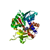
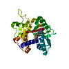
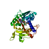
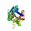
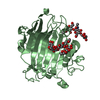
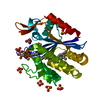
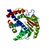
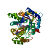
- PDB-1ri1: Structure and mechanism of mRNA cap (guanine N-7) methyltransferase -
+ Open data

- Basic information
| Entry | Database: PDB / ID: 1ri1 | ||||||
|---|---|---|---|---|---|---|---|
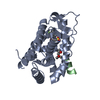
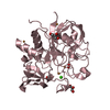
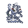
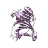
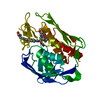
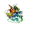
| Title | Structure and mechanism of mRNA cap (guanine N-7) methyltransferase | ||||||
 Components Components | mRNA CAPPING ENZYME | ||||||
 Keywords Keywords | TRANSFERASE / methyltransferase / rna / cap / m7G / messenger rna cap | ||||||
| Function / homology |  Function and homology information Function and homology informationmRNA (guanine-N7)-methyltransferase / mRNA 5'-cap (guanine-N7-)-methyltransferase activity / RNA binding / nucleus Similarity search - Function | ||||||
| Biological species |  Encephalitozoon cuniculi (fungus) Encephalitozoon cuniculi (fungus) | ||||||
| Method |  X-RAY DIFFRACTION / SYNCHROTRON / SAD, MIR / Resolution: 2.5 Å X-RAY DIFFRACTION / SYNCHROTRON / SAD, MIR / Resolution: 2.5 Å | ||||||
 Authors Authors | Fabrega, C. / Hausmann, S. / Shen, V. / Shuman, S. / Lima, C.D. | ||||||
 Citation Citation | Journal: Mol.Cell / Year: 2004 Title: Structure and mechanism of mRNA cap (Guanine-n7) methyltransferase Authors: Fabrega, C. / Hausmann, S. / Shen, V. / Shuman, S. / Lima, C.D. | ||||||
| History |
| ||||||
| Remark 600 | HETEROGEN 7mG and ribose are disordered leaving only a GTP modeled in the structure |
- Structure visualization
| Structure viewer | Molecule: MolmilJmol/JSmol |
|---|
- Downloads & links
-Download
| PDBx/mmCIF format | 1ri1.cif.gz | 67.9 KB | Display | PDBx/mmCIF format |
|---|---|---|---|---|
| PDB format | pdb1ri1.ent.gz | 49.2 KB | Display | PDB format |
| PDBx/mmJSON format | 1ri1.json.gz | Tree view | PDBx/mmJSON format | |
| Others |  Other downloads Other downloads |
-Validation report
| Arichive directory | https://data.pdbj.org/pub/pdb/validation_reports/ri/1ri1ftp://data.pdbj.org/pub/pdb/validation_reports/ri/1ri1 | HTTPS FTP |
|---|
-Related structure data
-Links
 PDBj
PDBj


- Assembly
| Deposited unit | 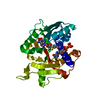
| ||||||||
|---|---|---|---|---|---|---|---|---|---|
| 1 |
| ||||||||
| Unit cell |
|
-Components
| #1: Protein | Mass: 34823.684 Da / Num. of mol.: 1 Source method: isolated from a genetically manipulated source Source: (gene. exp.) Encephalitozoon cuniculi (fungus) / Gene: ECU10_0380 / Plasmid: PSUMO-SMT3 / Species (production host): Escherichia coli / Production host:  |
|---|---|
| #2: Chemical | ChemComp-SAH /   Type: L-peptide linking / Mass: 384.411 Da / Num. of mol.: 1 / Source method: obtained synthetically / Formula: C14H20N6O5S Type: L-peptide linking / Mass: 384.411 Da / Num. of mol.: 1 / Source method: obtained synthetically / Formula: C14H20N6O5S |
| #3: Chemical | ChemComp-GTG /   Mass: 803.440 Da / Num. of mol.: 1 / Source method: obtained synthetically / Formula: C21H30N10O18P3 Mass: 803.440 Da / Num. of mol.: 1 / Source method: obtained synthetically / Formula: C21H30N10O18P3 |
| #4: Water | ChemComp-HOH /  Mass: 18.015 Da / Num. of mol.: 58 / Source method: isolated from a natural source / Formula: H2O Mass: 18.015 Da / Num. of mol.: 58 / Source method: isolated from a natural source / Formula: H2O |
-Experimental details
-Experiment
| Experiment | Method: X-RAY DIFFRACTION / Number of used crystals: 1 |
|---|
- Sample preparation
| Crystal | Density Matthews: 1.89 Å3/Da / Density % sol: 34.83 % | ||||||||||||||||||||||||||||||||||||||||||||||||||||||||
|---|---|---|---|---|---|---|---|---|---|---|---|---|---|---|---|---|---|---|---|---|---|---|---|---|---|---|---|---|---|---|---|---|---|---|---|---|---|---|---|---|---|---|---|---|---|---|---|---|---|---|---|---|---|---|---|---|---|
| Crystal grow | Temperature: 291 K / Method: vapor diffusion, hanging drop / pH: 6.25 Details: 1.2M Na/K tartrate, 50mM BIS/TRIS, 20mM DTT, pH 6.25, VAPOR DIFFUSION, HANGING DROP, temperature 291K | ||||||||||||||||||||||||||||||||||||||||||||||||||||||||
| Crystal grow | *PLUS pH: 8 / Method: vapor diffusion | ||||||||||||||||||||||||||||||||||||||||||||||||||||||||
| Components of the solutions | *PLUS
|
-Data collection
| Diffraction | Mean temperature: 100 K |
|---|---|
| Diffraction source | Source: SYNCHROTRON / Site: NSLS  / Beamline: X9A / Wavelength: 0.979 Å / Beamline: X9A / Wavelength: 0.979 Å |
| Detector | Type: MARRESEARCH / Detector: CCD |
| Radiation | Monochromator: Si / Protocol: SINGLE WAVELENGTH / Monochromatic (M) / Laue (L): M / Scattering type: x-ray |
| Radiation wavelength | Wavelength: 0.979 Å / Relative weight: 1 |
| Reflection | Resolution: 2.4→20 Å / Num. all: 10841 / Num. obs: 10451 / % possible obs: 96.4 % / Observed criterion σ(F): 0 / Observed criterion σ(I): 0 / Biso Wilson estimate: 47.1 Å2 / Rmerge(I) obs: 0.076 / Net I/σ(I): 16.1 |
| Reflection shell | Resolution: 2.4→2.5 Å / % possible all: 93.3 |
| Reflection | *PLUS Num. measured all: 139600 |
| Reflection shell | *PLUS % possible obs: 93.3 % / Rmerge(I) obs: 0.33 / Mean I/σ(I) obs: 3 |
- Processing
| Software |
| ||||||||||||||||||||||||||||||||||||
|---|---|---|---|---|---|---|---|---|---|---|---|---|---|---|---|---|---|---|---|---|---|---|---|---|---|---|---|---|---|---|---|---|---|---|---|---|---|
| Refinement | Method to determine structure: SAD, MIR / Resolution: 2.5→19.68 Å / Rfactor Rfree error: 0.013 / Data cutoff high absF: 1725937.45 / Data cutoff low absF: 0 / Isotropic thermal model: RESTRAINED / Cross valid method: THROUGHOUT / σ(F): 0 / Stereochemistry target values: Engh & Huber / Details: BULK SOLVENT MODEL USED
| ||||||||||||||||||||||||||||||||||||
| Solvent computation | Solvent model: FLAT MODEL / Bsol: 31.075 Å2 / ksol: 0.35419 e/Å3 | ||||||||||||||||||||||||||||||||||||
| Displacement parameters | Biso mean: 41.2 Å2
| ||||||||||||||||||||||||||||||||||||
| Refine analyze |
| ||||||||||||||||||||||||||||||||||||
| Refinement step | Cycle: LAST / Resolution: 2.5→19.68 Å
| ||||||||||||||||||||||||||||||||||||
| Refine LS restraints |
| ||||||||||||||||||||||||||||||||||||
| LS refinement shell | Resolution: 2.5→2.66 Å / Rfactor Rfree error: 0.039 / Total num. of bins used: 6
| ||||||||||||||||||||||||||||||||||||
| Xplor file |
| ||||||||||||||||||||||||||||||||||||
| Refinement | *PLUS Highest resolution: 2.5 Å / Lowest resolution: 20 Å / % reflection Rfree: 5 % / Rfactor Rfree: 0.28 / Rfactor Rwork: 0.22 | ||||||||||||||||||||||||||||||||||||
| Solvent computation | *PLUS | ||||||||||||||||||||||||||||||||||||
| Displacement parameters | *PLUS | ||||||||||||||||||||||||||||||||||||
| Refine LS restraints | *PLUS
| ||||||||||||||||||||||||||||||||||||
| LS refinement shell | *PLUS Rfactor Rfree: 0.36 / Rfactor Rwork: 0.31 |
