 Movie
Movie Controller

[English] 日本語
 Yorodumi
- PDB-2uyo: Crystal structure of ML2640c from Mycobacterium leprae in an hexa... -
+ Open data

- Basic information
| Entry | Database: PDB / ID: 2uyo | ||||||
|---|---|---|---|---|---|---|---|
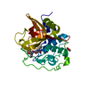
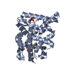
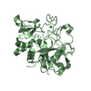
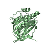
| Title | Crystal structure of ML2640c from Mycobacterium leprae in an hexagonal crystal form | ||||||
 Components Components | HYPOTHETICAL PROTEIN ML2640 | ||||||
 Keywords Keywords | TRANSFERASE / HYPOTHETICAL PROTEIN / PUTATIVE METHYLTRANSFERASE | ||||||
| Function / homology |  Function and homology information Function and homology informationTransferases; Transferring one-carbon groups; Methyltransferases / methyltransferase activity / methylation Similarity search - Function | ||||||
| Biological species |  MYCOBACTERIUM LEPRAE (bacteria) MYCOBACTERIUM LEPRAE (bacteria) | ||||||
| Method |  X-RAY DIFFRACTION / SYNCHROTRON / MOLECULAR REPLACEMENT / Resolution: 1.7 Å X-RAY DIFFRACTION / SYNCHROTRON / MOLECULAR REPLACEMENT / Resolution: 1.7 Å | ||||||
 Authors Authors | Grana, M. / Buschiazzo, A. / Wehenkel, A. / Haouz, A. / Miras, I. / Shepard, W. / Alzari, P.M. | ||||||
 Citation Citation | Journal: Protein Sci. / Year: 2007 Title: The Crystal Structure of M. Leprae Ml2640C Defines a Large Family of Putative S-Adenosylmethionine- Dependent Methyltransferases in Mycobacteria. Authors: Grana, M. / Haouz, A. / Buschiazzo, A. / Miras, I. / Wehenkel, A. / Bondet, V. / Shepard, W. / Schaeffer, F. / Cole, S.T. / Alzari, P.M. | ||||||
| History |
|
- Structure visualization
| Structure viewer | Molecule: MolmilJmol/JSmol |
|---|
- Downloads & links
-Download
| PDBx/mmCIF format | 2uyo.cif.gz | 70.2 KB | Display | PDBx/mmCIF format |
|---|---|---|---|---|
| PDB format | pdb2uyo.ent.gz | 50.6 KB | Display | PDB format |
| PDBx/mmJSON format | 2uyo.json.gz | Tree view | PDBx/mmJSON format | |
| Others |  Other downloads Other downloads |
-Validation report
| Arichive directory | https://data.pdbj.org/pub/pdb/validation_reports/uy/2uyoftp://data.pdbj.org/pub/pdb/validation_reports/uy/2uyo | HTTPS FTP |
|---|
-Related structure data
| Related structure data |  2ckdSC  2uyqC S: Starting model for refinement C: citing same article ( |
|---|---|
| Similar structure data |

-Links
 PDBj
PDBj- Assembly
| Deposited unit | 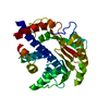
| ||||||||
|---|---|---|---|---|---|---|---|---|---|
| 1 |
| ||||||||
| Unit cell |
|
-Components
| #1: Protein | Mass: 34492.656 Da / Num. of mol.: 1 Source method: isolated from a genetically manipulated source Source: (gene. exp.) MYCOBACTERIUM LEPRAE (bacteria) / Production host: |
|---|---|
| #2: Water | ChemComp-HOH /  Mass: 18.015 Da / Num. of mol.: 211 / Source method: isolated from a natural source / Formula: H2O Mass: 18.015 Da / Num. of mol.: 211 / Source method: isolated from a natural source / Formula: H2O |
-Experimental details
-Experiment
| Experiment | Method: X-RAY DIFFRACTION / Number of used crystals: 1 |
|---|
- Sample preparation
| Crystal | Density Matthews: 2.5 Å3/Da / Density % sol: 51 % / Description: NONE |
|---|---|
| Crystal grow | pH: 9 / Details: 0.8 M SODIUM CITRATE, 100 MM BICINE, PH 9 |
-Data collection
| Diffraction | Mean temperature: 100 K |
|---|---|
| Diffraction source | Source: SYNCHROTRON / Site: ESRF  / Beamline: ID29 / Wavelength: 0.9756 / Beamline: ID29 / Wavelength: 0.9756 |
| Detector | Type: ADSC CCD / Detector: CCD / Date: Feb 7, 2005 |
| Radiation | Protocol: SINGLE WAVELENGTH / Monochromatic (M) / Laue (L): M / Scattering type: x-ray |
| Radiation wavelength | Wavelength: 0.9756 Å / Relative weight: 1 |
| Reflection | Resolution: 1.7→50 Å / Num. obs: 37485 / % possible obs: 99.3 % / Observed criterion σ(I): 0 / Redundancy: 6.8 % / Rmerge(I) obs: 0.09 / Net I/σ(I): 4.3 |
| Reflection shell | Resolution: 1.7→1.79 Å / Redundancy: 5 % / Rmerge(I) obs: 0.41 / Mean I/σ(I) obs: 1.6 / % possible all: 99.9 |
- Processing
| Software |
| ||||||||||||||||||||||||||||||||||||||||||||||||||||||||||||||||||||||||||||||||||||||||||||||||||||||||||||||||||||||||||||||||||||||||||||||||||||||||||||||||||||||||||||||||||||||
|---|---|---|---|---|---|---|---|---|---|---|---|---|---|---|---|---|---|---|---|---|---|---|---|---|---|---|---|---|---|---|---|---|---|---|---|---|---|---|---|---|---|---|---|---|---|---|---|---|---|---|---|---|---|---|---|---|---|---|---|---|---|---|---|---|---|---|---|---|---|---|---|---|---|---|---|---|---|---|---|---|---|---|---|---|---|---|---|---|---|---|---|---|---|---|---|---|---|---|---|---|---|---|---|---|---|---|---|---|---|---|---|---|---|---|---|---|---|---|---|---|---|---|---|---|---|---|---|---|---|---|---|---|---|---|---|---|---|---|---|---|---|---|---|---|---|---|---|---|---|---|---|---|---|---|---|---|---|---|---|---|---|---|---|---|---|---|---|---|---|---|---|---|---|---|---|---|---|---|---|---|---|---|---|
| Refinement | Method to determine structure: MOLECULAR REPLACEMENT Starting model: PDB ENTRY 2CKD Resolution: 1.7→65.51 Å / Cor.coef. Fo:Fc: 0.946 / Cor.coef. Fo:Fc free: 0.932 / SU B: 4.203 / SU ML: 0.072 / TLS residual ADP flag: LIKELY RESIDUAL / Cross valid method: THROUGHOUT / ESU R: 0.105 / ESU R Free: 0.102 / Stereochemistry target values: MAXIMUM LIKELIHOOD / Details: HYDROGENS HAVE BEEN ADDED IN THE RIDING POSITIONS.
| ||||||||||||||||||||||||||||||||||||||||||||||||||||||||||||||||||||||||||||||||||||||||||||||||||||||||||||||||||||||||||||||||||||||||||||||||||||||||||||||||||||||||||||||||||||||
| Solvent computation | Ion probe radii: 0.8 Å / Shrinkage radii: 0.8 Å / VDW probe radii: 1.2 Å / Solvent model: MASK | ||||||||||||||||||||||||||||||||||||||||||||||||||||||||||||||||||||||||||||||||||||||||||||||||||||||||||||||||||||||||||||||||||||||||||||||||||||||||||||||||||||||||||||||||||||||
| Displacement parameters | Biso mean: 28.72 Å2
| ||||||||||||||||||||||||||||||||||||||||||||||||||||||||||||||||||||||||||||||||||||||||||||||||||||||||||||||||||||||||||||||||||||||||||||||||||||||||||||||||||||||||||||||||||||||
| Refinement step | Cycle: LAST / Resolution: 1.7→65.51 Å
| ||||||||||||||||||||||||||||||||||||||||||||||||||||||||||||||||||||||||||||||||||||||||||||||||||||||||||||||||||||||||||||||||||||||||||||||||||||||||||||||||||||||||||||||||||||||
| Refine LS restraints |
|