 Movie
Movie Controller

[English] 日本語
 Yorodumi
- PDB-7d7q: Crystal structure of the transmembrane domain and linker region o... -
+ Open data

- Basic information
| Entry | Database: PDB / ID: 7d7q | ||||||
|---|---|---|---|---|---|---|---|
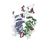
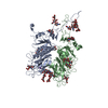
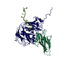
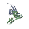
| Title | Crystal structure of the transmembrane domain and linker region of Salpingoeca rosetta rhodopsin phosphodiesterase | ||||||
 Components Components | Phosphodiesterase | ||||||
 Keywords Keywords | MEMBRANE PROTEIN / microbial rhodopsin / eight-transmembrane / light-dependent phosphodiesterase | ||||||
| Function / homology |  Function and homology information Function and homology informationHydrolases; Acting on ester bonds; Phosphoric-diester hydrolases / 3',5'-cyclic-nucleotide phosphodiesterase activity / signal transduction / membrane / metal ion binding Similarity search - Function | ||||||
| Biological species |  Salpingoeca rosetta (eukaryote) Salpingoeca rosetta (eukaryote) | ||||||
| Method |  X-RAY DIFFRACTION / SYNCHROTRON / MOLECULAR REPLACEMENT / Resolution: 3.5 Å X-RAY DIFFRACTION / SYNCHROTRON / MOLECULAR REPLACEMENT / Resolution: 3.5 Å | ||||||
 Authors Authors | Ikuta, T. / Shihoya, W. / Yamashita, K. / Nureki, O. | ||||||
 Citation Citation | Journal: Nat Commun / Year: 2020 Title: Structural insights into the mechanism of rhodopsin phosphodiesterase. Authors: Ikuta, T. / Shihoya, W. / Sugiura, M. / Yoshida, K. / Watari, M. / Tokano, T. / Yamashita, K. / Katayama, K. / Tsunoda, S.P. / Uchihashi, T. / Kandori, H. / Nureki, O. | ||||||
| History |
|
- Structure visualization
| Structure viewer | Molecule: MolmilJmol/JSmol |
|---|
- Downloads & links
-Download
| PDBx/mmCIF format | 7d7q.cif.gz | 156.3 KB | Display | PDBx/mmCIF format |
|---|---|---|---|---|
| PDB format | pdb7d7q.ent.gz | 98.8 KB | Display | PDB format |
| PDBx/mmJSON format | 7d7q.json.gz | Tree view | PDBx/mmJSON format | |
| Others |  Other downloads Other downloads |
-Validation report
| Arichive directory | https://data.pdbj.org/pub/pdb/validation_reports/d7/7d7qftp://data.pdbj.org/pub/pdb/validation_reports/d7/7d7q | HTTPS FTP |
|---|
-Related structure data
| Related structure data | 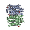 7cj3SC  7d7pC S: Starting model for refinement C: citing same article ( |
|---|---|
| Similar structure data | |
| Experimental dataset #1 | Data reference: 10.5281/zenodo.4080668 / Data set type: diffraction image data |


-Links
 PDBj
PDBj


- Assembly
| Deposited unit | 
| ||||||||||||
|---|---|---|---|---|---|---|---|---|---|---|---|---|---|
| 1 |
| ||||||||||||
| Unit cell |
|
-Components
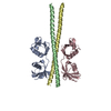
| #1: Protein | Mass: 39263.566 Da / Num. of mol.: 2 Source method: isolated from a genetically manipulated source Source: (gene. exp.) Salpingoeca rosetta (strain ATCC 50818 / BSB-021) (eukaryote)Gene: PTSG_02023 / Production host:  Homo sapiens (human) Homo sapiens (human)References: UniProt: F2TZN0, Hydrolases; Acting on ester bonds; Phosphoric-diester hydrolases #2: Chemical |   Mass: 284.436 Da / Num. of mol.: 2 / Source method: obtained synthetically / Formula: C20H28O Mass: 284.436 Da / Num. of mol.: 2 / Source method: obtained synthetically / Formula: C20H28O#3: Chemical | ChemComp-OLC / (   Mass: 356.540 Da / Num. of mol.: 4 / Source method: obtained synthetically / Formula: C21H40O4 Mass: 356.540 Da / Num. of mol.: 4 / Source method: obtained synthetically / Formula: C21H40O4Has ligand of interest | N | Has protein modification | Y | |
|---|
-Experimental details
-Experiment
| Experiment | Method: X-RAY DIFFRACTION / Number of used crystals: 1 |
|---|
- Sample preparation
| Crystal | Density Matthews: 3.42 Å3/Da / Density % sol: 64.09 % |
|---|---|
| Crystal grow | Temperature: 293 K / Method: lipidic cubic phase / pH: 5 Details: 25% (w/v) PEG 500DME, 100mM Na-citrate, pH 5.0, 100mM NaK-tartrate |
-Data collection
| Diffraction | Mean temperature: 100 K / Serial crystal experiment: N |
|---|---|
| Diffraction source | Source: SYNCHROTRON / Site: SPring-8  / Beamline: BL32XU / Wavelength: 1 Å / Beamline: BL32XU / Wavelength: 1 Å |
| Detector | Type: DECTRIS EIGER X 9M / Detector: PIXEL / Date: Apr 10, 2019 |
| Radiation | Protocol: SINGLE WAVELENGTH / Monochromatic (M) / Laue (L): M / Scattering type: x-ray |
| Radiation wavelength | Wavelength: 1 Å / Relative weight: 1 |
| Reflection | Resolution: 3.5→49.4 Å / Num. obs: 13712 / % possible obs: 98.1 % / Redundancy: 6.55 % / Biso Wilson estimate: 49.61 Å2 / CC1/2: 0.934 / Rrim(I) all: 0.673 / Net I/σ(I): 3.9 |
| Reflection shell | Resolution: 3.5→3.71 Å / Num. unique obs: 2147 / CC1/2: 0.393 / Rrim(I) all: 2.868 |
- Processing
| Software |
| ||||||||||||||||||||||||||||||||||||||||||
|---|---|---|---|---|---|---|---|---|---|---|---|---|---|---|---|---|---|---|---|---|---|---|---|---|---|---|---|---|---|---|---|---|---|---|---|---|---|---|---|---|---|---|---|
| Refinement | Method to determine structure: MOLECULAR REPLACEMENT Starting model: 7CJ3 Resolution: 3.5→49.4 Å / SU ML: 0.4813 / Cross valid method: FREE R-VALUE / σ(F): 1.34 / Phase error: 29.3612 Stereochemistry target values: GeoStd + Monomer Library + CDL v1.2
| ||||||||||||||||||||||||||||||||||||||||||
| Solvent computation | Shrinkage radii: 0.9 Å / VDW probe radii: 1.11 Å / Solvent model: FLAT BULK SOLVENT MODEL | ||||||||||||||||||||||||||||||||||||||||||
| Displacement parameters | Biso mean: 33.37 Å2 | ||||||||||||||||||||||||||||||||||||||||||
| Refinement step | Cycle: LAST / Resolution: 3.5→49.4 Å
| ||||||||||||||||||||||||||||||||||||||||||
| Refine LS restraints |
| ||||||||||||||||||||||||||||||||||||||||||
| LS refinement shell |
|
