 Movie
Movie Controller

[English] 日本語
 Yorodumi
- PDB-7d6z: Molecular model of the cryo-EM structure of 70S ribosome in compl... -
+ Open data

- Basic information
| Entry | Database: PDB / ID: 7d6z | ||||||||||||||||||||||||||||||||||||
|---|---|---|---|---|---|---|---|---|---|---|---|---|---|---|---|---|---|---|---|---|---|---|---|---|---|---|---|---|---|---|---|---|---|---|---|---|---|
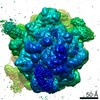
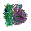
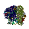
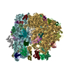
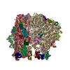
| Title | Molecular model of the cryo-EM structure of 70S ribosome in complex with peptide deformylase and trigger factor | ||||||||||||||||||||||||||||||||||||
 Components Components |
| ||||||||||||||||||||||||||||||||||||
 Keywords Keywords | RIBOSOME / Escherichia coli / nascent chain / protein biogenesis / peptide deformylase / trigger factor | ||||||||||||||||||||||||||||||||||||
| Function / homology |  Function and homology information Function and homology informationN-terminal protein amino acid modification / 'de novo' cotranslational protein folding / stress response to copper ion / peptide deformylase / peptide deformylase activity / stringent response / negative regulation of cytoplasmic translational initiation / transcription antitermination factor activity, RNA binding / ornithine decarboxylase inhibitor activity / misfolded RNA binding ...N-terminal protein amino acid modification / 'de novo' cotranslational protein folding / stress response to copper ion / peptide deformylase / peptide deformylase activity / stringent response / negative regulation of cytoplasmic translational initiation / transcription antitermination factor activity, RNA binding / ornithine decarboxylase inhibitor activity / misfolded RNA binding / Group I intron splicing / protein unfolding / RNA folding / translational termination / transcriptional attenuation / endoribonuclease inhibitor activity / positive regulation of ribosome biogenesis / RNA-binding transcription regulator activity / four-way junction DNA binding / negative regulation of cytoplasmic translation / regulation of mRNA stability / DnaA-L2 complex / translation repressor activity / protein folding chaperone / negative regulation of translational initiation / negative regulation of DNA-templated DNA replication initiation / mRNA regulatory element binding translation repressor activity / positive regulation of RNA splicing / regulation of DNA-templated transcription elongation / response to reactive oxygen species / transcription elongation factor complex / cytosolic ribosome assembly / ribosome assembly / peptidylprolyl isomerase / assembly of large subunit precursor of preribosome / peptidyl-prolyl cis-trans isomerase activity / transcription antitermination / DNA endonuclease activity / regulation of cell growth / translational initiation / DNA-templated transcription termination / ferrous iron binding / response to radiation / maintenance of translational fidelity / mRNA 5'-UTR binding / regulation of translation / protein transport / large ribosomal subunit / response to heat / transferase activity / ribosomal small subunit assembly / ribosome binding / ribosome biogenesis / ribosomal small subunit biogenesis / protein folding / 5S rRNA binding / ribosomal large subunit assembly / small ribosomal subunit / small ribosomal subunit rRNA binding / cytosolic small ribosomal subunit / large ribosomal subunit rRNA binding / cytosolic large ribosomal subunit / cytoplasmic translation / tRNA binding / negative regulation of translation / rRNA binding / structural constituent of ribosome / ribosome / translation / cell division / response to antibiotic / negative regulation of DNA-templated transcription / hydrolase activity / mRNA binding / DNA binding / RNA binding / zinc ion binding / membrane / identical protein binding / cytosol / cytoplasm Similarity search - Function | ||||||||||||||||||||||||||||||||||||
| Biological species |  | ||||||||||||||||||||||||||||||||||||
| Method | ELECTRON MICROSCOPY / single particle reconstruction / cryo EM / Resolution: 3.4 Å | ||||||||||||||||||||||||||||||||||||
 Authors Authors | Akbar, S. / Bhakta, S. / Sengupta, J. | ||||||||||||||||||||||||||||||||||||
| Funding support |  India, 3items India, 3items
| ||||||||||||||||||||||||||||||||||||
 Citation Citation | Journal: Structure / Year: 2021 Title: Structural insights into the interplay of protein biogenesis factors with the 70S ribosome. Authors: Shirin Akbar / Sayan Bhakta / Jayati Sengupta / Abstract: Bacterial co-translational N-terminal methionine excision, an early event of nascent polypeptide chain processing, is mediated by two enzymes: peptide deformylase (PDF) and methionine aminopeptidase ...Bacterial co-translational N-terminal methionine excision, an early event of nascent polypeptide chain processing, is mediated by two enzymes: peptide deformylase (PDF) and methionine aminopeptidase (MetAP). Trigger factor (TF), the only ribosome-associated bacterial chaperone, offers co-translational chaperoning assistance. Here, we present two high-resolution cryoelectron microscopy structures of tRNA-bound E. coli ribosome complexes showing simultaneous binding of PDF and TF, in the absence (3.4 Å) and presence of MetAP (4.1 Å). These structures establish molecular details of the interactions of the factors with the ribosome, and thereby reveal the structural basis of nascent chain processing. Our results suggest that simultaneous binding of all three factors is not a functionally favorable mechanism of nascent chain processing. Strikingly, an unusual structural distortion of the 70S ribosome, potentially driven by binding of multiple copies of MetAP, is observed when MetAP is incubated with a pre-formed PDF-TF-bound ribosome complex. | ||||||||||||||||||||||||||||||||||||
| History |
|
- Structure visualization
| Movie |
Movie viewer |
|---|---|
| Structure viewer | Molecule: MolmilJmol/JSmol |
- Downloads & links
-Download
| PDBx/mmCIF format | 7d6z.cif.gz | 3.2 MB | Display | PDBx/mmCIF format |
|---|---|---|---|---|
| PDB format | pdb7d6z.ent.gz | Display | PDB format | |
| PDBx/mmJSON format | 7d6z.json.gz | Tree view | PDBx/mmJSON format | |
| Others |  Other downloads Other downloads |
-Validation report
| Arichive directory | https://data.pdbj.org/pub/pdb/validation_reports/d6/7d6zftp://data.pdbj.org/pub/pdb/validation_reports/d6/7d6z | HTTPS FTP |
|---|
-Related structure data
| Related structure data | 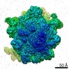 30598MC  7d80C M: map data used to model this data C: citing same article ( |
|---|---|
| Similar structure data |


-Links
 PDBj
PDBj


- Assembly
| Deposited unit | 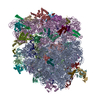
|
|---|---|
| 1 |
|
-Components
-30S ribosomal protein ... , 20 types, 20 molecules 01ijklmnopqrstuvwxyz
| #1: Protein | Mass: 9708.464 Da / Num. of mol.: 1 / Source method: isolated from a natural source / Source: (natural) |
|---|---|
| #2: Protein | Mass: 8524.039 Da / Num. of mol.: 1 / Source method: isolated from a natural source / Source: (natural) |
| #41: Protein | Mass: 26781.670 Da / Num. of mol.: 1 / Source method: isolated from a natural source / Source: (natural) |
| #42: Protein | Mass: 26031.316 Da / Num. of mol.: 1 / Source method: isolated from a natural source / Source: (natural) |
| #43: Protein | Mass: 23514.199 Da / Num. of mol.: 1 / Source method: isolated from a natural source / Source: (natural) |
| #44: Protein | Mass: 17629.398 Da / Num. of mol.: 1 / Source method: isolated from a natural source / Source: (natural) |
| #45: Protein | Mass: 15727.512 Da / Num. of mol.: 1 / Source method: isolated from a natural source / Source: (natural) |
| #46: Protein | Mass: 20055.156 Da / Num. of mol.: 1 / Source method: isolated from a natural source / Source: (natural) |
| #47: Protein | Mass: 14146.557 Da / Num. of mol.: 1 / Source method: isolated from a natural source / Source: (natural) |
| #48: Protein | Mass: 14886.270 Da / Num. of mol.: 1 / Source method: isolated from a natural source / Source: (natural) |
| #49: Protein | Mass: 11755.597 Da / Num. of mol.: 1 / Source method: isolated from a natural source / Source: (natural) |
| #50: Protein | Mass: 13870.975 Da / Num. of mol.: 1 / Source method: isolated from a natural source / Source: (natural) |
| #51: Protein | Mass: 13768.157 Da / Num. of mol.: 1 / Source method: isolated from a natural source / Source: (natural) |
| #52: Protein | Mass: 13128.467 Da / Num. of mol.: 1 / Source method: isolated from a natural source / Source: (natural) |
| #53: Protein | Mass: 11606.560 Da / Num. of mol.: 1 / Source method: isolated from a natural source / Source: (natural) |
| #54: Protein | Mass: 10290.816 Da / Num. of mol.: 1 / Source method: isolated from a natural source / Source: (natural) |
| #55: Protein | Mass: 9207.572 Da / Num. of mol.: 1 / Source method: isolated from a natural source / Source: (natural) |
| #56: Protein | Mass: 9724.491 Da / Num. of mol.: 1 / Source method: isolated from a natural source / Source: (natural) |
| #57: Protein | Mass: 9005.472 Da / Num. of mol.: 1 / Source method: isolated from a natural source / Source: (natural) |
| #58: Protein | Mass: 10455.355 Da / Num. of mol.: 1 / Source method: isolated from a natural source / Source: (natural) |
-RNA chain , 6 types, 6 molecules 234ABf
| #3: RNA chain | Mass: 23678.199 Da / Num. of mol.: 1 / Source method: isolated from a natural source / Source: (natural) |
|---|---|
| #4: RNA chain | Mass: 25004.014 Da / Num. of mol.: 1 / Source method: isolated from a natural source / Source: (natural) |
| #5: RNA chain | Mass: 24728.910 Da / Num. of mol.: 1 / Source method: isolated from a natural source / Source: (natural) |
| #7: RNA chain | Mass: 941306.188 Da / Num. of mol.: 1 / Source method: isolated from a natural source / Source: (natural) |
| #8: RNA chain | Mass: 38177.762 Da / Num. of mol.: 1 / Source method: isolated from a natural source / Source: (natural) |
| #38: RNA chain | Mass: 498725.406 Da / Num. of mol.: 1 / Source method: isolated from a natural source / Source: (natural) |
+50S ribosomal protein ... , 30 types, 30 molecules 6CDEFGHIJKLMNOPQRSTUVWXYZabcde
-Protein , 2 types, 2 molecules gh
| #39: Protein | Mass: 19357.447 Da / Num. of mol.: 1 Source method: isolated from a genetically manipulated source Source: (gene. exp.) Gene: def, fms, b3287, JW3248 / Production host: |
|---|---|
| #40: Protein | Mass: 13091.964 Da / Num. of mol.: 1 Source method: isolated from a genetically manipulated source Source: (gene. exp.) Gene: tig, b0436, JW0426 / Production host: |
-Details
| Has protein modification | Y |
|---|---|
| Source details | Purified tRNAs were purchased from Sigma, and ribosomes were purified from E. coli MRE600 strain. ...Purified tRNAs were purchased from Sigma, and ribosomes were purified from E. coli MRE600 strain. And non-ribosomal proteins peptide deformylase and tigger factor are from E. coli (strain K12). |
-Experimental details
-Experiment
| Experiment | Method: ELECTRON MICROSCOPY |
|---|---|
| EM experiment | Aggregation state: PARTICLE / 3D reconstruction method: single particle reconstruction |
- Sample preparation
| Component | Name: E. coli 70S ribosome in complex with enzyme peptide deformylase and chaperone trigger factor Type: RIBOSOME / Entity ID: all / Source: MULTIPLE SOURCES |
|---|---|
| Source (natural) | Organism: |
| Buffer solution | pH: 7.4 |
| Specimen | Embedding applied: NO / Shadowing applied: NO / Staining applied: NO / Vitrification applied: YES |
| Specimen support | Grid material: COPPER / Grid mesh size: 300 divisions/in. / Grid type: Quantifoil |
| Vitrification | Instrument: FEI VITROBOT MARK IV / Cryogen name: ETHANE / Humidity: 100 % |
- Electron microscopy imaging
| Experimental equipment |  Model: Titan Krios / Image courtesy: FEI Company |
|---|---|
| Microscopy | Model: FEI TITAN KRIOS |
| Electron gun | Electron source:  FIELD EMISSION GUN / Accelerating voltage: 300 kV / Illumination mode: FLOOD BEAM FIELD EMISSION GUN / Accelerating voltage: 300 kV / Illumination mode: FLOOD BEAM |
| Electron lens | Mode: BRIGHT FIELD |
| Image recording | Electron dose: 32.57 e/Å2 / Film or detector model: FEI FALCON III (4k x 4k) |
- Processing
| EM software |
| ||||||||||||||||||||||||||||||||
|---|---|---|---|---|---|---|---|---|---|---|---|---|---|---|---|---|---|---|---|---|---|---|---|---|---|---|---|---|---|---|---|---|---|
| CTF correction | Type: PHASE FLIPPING AND AMPLITUDE CORRECTION | ||||||||||||||||||||||||||||||||
| Symmetry | Point symmetry: C1 (asymmetric) | ||||||||||||||||||||||||||||||||
| 3D reconstruction | Resolution: 3.4 Å / Resolution method: FSC 0.143 CUT-OFF / Num. of particles: 194157 / Symmetry type: POINT |
