 Movie
Movie Controller


+ Open data

- Basic information
| Entry | Database: PDB / ID: 7d53 | |||||||||
|---|---|---|---|---|---|---|---|---|---|---|
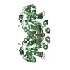
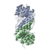
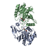
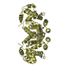
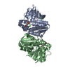
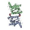
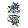
| Title | SpuA mutant - H221N with Glu | |||||||||
 Components Components | Probable glutamine amidotransferase | |||||||||
 Keywords Keywords | HYDROLASE / SpuA / gama-glutamyl-gama-aminobutyrate hydrolase / glutamine amidotransferase / Pseudomonas aeruginosa PAO1. | |||||||||
| Function / homology |  Function and homology information Function and homology informationgamma-glutamyl-gamma-aminobutyrate hydrolase / gamma-glutamyl-gamma-aminobutyrate hydrolase activity / polyamine catabolic process / spermidine catabolic process / metal ion binding Similarity search - Function | |||||||||
| Biological species |  Pseudomonas aeruginosa PAO1 (bacteria) Pseudomonas aeruginosa PAO1 (bacteria) | |||||||||
| Method |  X-RAY DIFFRACTION / SYNCHROTRON / MOLECULAR REPLACEMENT / Resolution: 1.6 Å X-RAY DIFFRACTION / SYNCHROTRON / MOLECULAR REPLACEMENT / Resolution: 1.6 Å | |||||||||
 Authors Authors | Chen, Y. / Zhang, Q. / Bartlam, M. | |||||||||
| Funding support |  China, 2items China, 2items
| |||||||||
 Citation Citation | Journal: Acta Crystallogr D Struct Biol / Year: 2021 Title: Structure and mechanism of the gamma-glutamyl-gamma-aminobutyrate hydrolase SpuA from Pseudomonas aeruginosa. Authors: Chen, Y. / Jia, H. / Zhang, J. / Liang, Y. / Liu, R. / Zhang, Q. / Bartlam, M. | |||||||||
| History |
|
- Structure visualization
| Structure viewer | Molecule: MolmilJmol/JSmol |
|---|
- Downloads & links
-Download
| PDBx/mmCIF format | 7d53.cif.gz | 456.6 KB | Display | PDBx/mmCIF format |
|---|---|---|---|---|
| PDB format | pdb7d53.ent.gz | 301.7 KB | Display | PDB format |
| PDBx/mmJSON format | 7d53.json.gz | Tree view | PDBx/mmJSON format | |
| Others |  Other downloads Other downloads |
-Validation report
| Arichive directory | https://data.pdbj.org/pub/pdb/validation_reports/d5/7d53ftp://data.pdbj.org/pub/pdb/validation_reports/d5/7d53 | HTTPS FTP |
|---|
-Related structure data
| Related structure data | 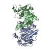 7d4rSC  7d50C 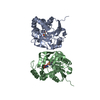 7d54C S: Starting model for refinement C: citing same article ( |
|---|---|
| Similar structure data |


-Links
 PDBj
PDBj


- Assembly
| Deposited unit | 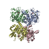
| ||||||||||||
|---|---|---|---|---|---|---|---|---|---|---|---|---|---|
| 1 | 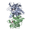
| ||||||||||||
| 2 | 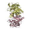
| ||||||||||||
| Unit cell |
|
-Components
| #1: Protein | Mass: 26482.100 Da / Num. of mol.: 4 / Mutation: H221N Source method: isolated from a genetically manipulated source Source: (gene. exp.) Pseudomonas aeruginosa PAO1 (bacteria) / Strain: PAO1 / Gene: spuA, PA0297 / Production host: #2: Chemical | ChemComp-MG /   Mass: 24.305 Da / Num. of mol.: 4 / Source method: obtained synthetically / Formula: Mg / Feature type: SUBJECT OF INVESTIGATION Mass: 24.305 Da / Num. of mol.: 4 / Source method: obtained synthetically / Formula: Mg / Feature type: SUBJECT OF INVESTIGATION#3: Chemical | ChemComp-GLU /   Type: L-peptide linking / Mass: 147.129 Da / Num. of mol.: 4 / Source method: obtained synthetically / Formula: C5H9NO4 Type: L-peptide linking / Mass: 147.129 Da / Num. of mol.: 4 / Source method: obtained synthetically / Formula: C5H9NO4#4: Water | ChemComp-HOH / |  Mass: 18.015 Da / Num. of mol.: 1006 / Source method: isolated from a natural source / Formula: H2O Mass: 18.015 Da / Num. of mol.: 1006 / Source method: isolated from a natural source / Formula: H2OHas ligand of interest | Y | |
|---|
-Experimental details
-Experiment
| Experiment | Method: X-RAY DIFFRACTION / Number of used crystals: 1 |
|---|
- Sample preparation
| Crystal | Density Matthews: 2.13 Å3/Da / Density % sol: 42.13 % |
|---|---|
| Crystal grow | Temperature: 293 K / Method: vapor diffusion / pH: 8.5 Details: 0.2M Magnesium chloride hexahydrate, 0.1M Tris-HCl pH 8.5, 30% w/v Polyethylene glycol 4,000 |
-Data collection
| Diffraction | Mean temperature: 100 K / Serial crystal experiment: N |
|---|---|
| Diffraction source | Source: SYNCHROTRON / Site: SSRF / Beamline: BL18U1 / Wavelength: 0.97853 Å |
| Detector | Type: DECTRIS PILATUS3 6M / Detector: PIXEL / Date: Nov 9, 2019 |
| Radiation | Protocol: SINGLE WAVELENGTH / Monochromatic (M) / Laue (L): M / Scattering type: x-ray |
| Radiation wavelength | Wavelength: 0.97853 Å / Relative weight: 1 |
| Reflection | Resolution: 1.6→50 Å / Num. obs: 120939 / % possible obs: 100 % / Redundancy: 6.4 % / Biso Wilson estimate: 22.93 Å2 / Rpim(I) all: 0.04 / Net I/σ(I): 27.7 |
| Reflection shell | Resolution: 1.6→1.63 Å / Redundancy: 5.9 % / Mean I/σ(I) obs: 2 / Num. unique obs: 5983 / Rpim(I) all: 0.359 / % possible all: 100 |
- Processing
| Software |
| |||||||||||||||||||||||||||||||||||||||||||||||||||||||||||||||||||||||||||||||||||||||||||||||||||||||||
|---|---|---|---|---|---|---|---|---|---|---|---|---|---|---|---|---|---|---|---|---|---|---|---|---|---|---|---|---|---|---|---|---|---|---|---|---|---|---|---|---|---|---|---|---|---|---|---|---|---|---|---|---|---|---|---|---|---|---|---|---|---|---|---|---|---|---|---|---|---|---|---|---|---|---|---|---|---|---|---|---|---|---|---|---|---|---|---|---|---|---|---|---|---|---|---|---|---|---|---|---|---|---|---|---|---|---|
| Refinement | Method to determine structure: MOLECULAR REPLACEMENT Starting model: 7D4R Resolution: 1.6→30.61 Å / SU ML: 0.1507 / Cross valid method: FREE R-VALUE / σ(F): 1.36 / Phase error: 19.7517 Stereochemistry target values: GeoStd + Monomer Library + CDL v1.2
| |||||||||||||||||||||||||||||||||||||||||||||||||||||||||||||||||||||||||||||||||||||||||||||||||||||||||
| Solvent computation | Shrinkage radii: 0.9 Å / VDW probe radii: 1.11 Å / Solvent model: FLAT BULK SOLVENT MODEL | |||||||||||||||||||||||||||||||||||||||||||||||||||||||||||||||||||||||||||||||||||||||||||||||||||||||||
| Displacement parameters | Biso mean: 29.86 Å2 | |||||||||||||||||||||||||||||||||||||||||||||||||||||||||||||||||||||||||||||||||||||||||||||||||||||||||
| Refinement step | Cycle: LAST / Resolution: 1.6→30.61 Å
| |||||||||||||||||||||||||||||||||||||||||||||||||||||||||||||||||||||||||||||||||||||||||||||||||||||||||
| Refine LS restraints |
| |||||||||||||||||||||||||||||||||||||||||||||||||||||||||||||||||||||||||||||||||||||||||||||||||||||||||
| LS refinement shell |
|
