 Movie
Movie Controller

[English] 日本語
 Yorodumi
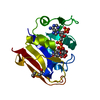
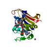
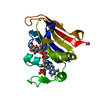
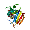
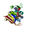
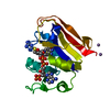
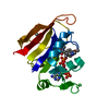
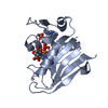
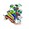
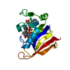
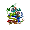
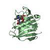
- PDB-7d4l: X-ray crystal Structure of E.coli Dihydrofolate Reductase complex... -
+ Open data

- Basic information
| Entry | Database: PDB / ID: 7d4l | |||||||||
|---|---|---|---|---|---|---|---|---|---|---|
| Title | X-ray crystal Structure of E.coli Dihydrofolate Reductase complexed with folate and NADP+ at pH7.0 | |||||||||
 Components Components | Dihydrofolate reductase | |||||||||
 Keywords Keywords | OXIDOREDUCTASE / dihydrofolate / reductase / alpha-beta sandiwitch / NADPH | |||||||||
| Function / homology |  Function and homology information Function and homology informationmethotrexate binding / dihydrofolic acid binding / 10-formyltetrahydrofolate biosynthetic process / response to methotrexate / folic acid biosynthetic process / folic acid binding / NADP+ binding / dihydrofolate metabolic process / dihydrofolate reductase / dihydrofolate reductase activity ...methotrexate binding / dihydrofolic acid binding / 10-formyltetrahydrofolate biosynthetic process / response to methotrexate / folic acid biosynthetic process / folic acid binding / NADP+ binding / dihydrofolate metabolic process / dihydrofolate reductase / dihydrofolate reductase activity / folic acid metabolic process / tetrahydrofolate biosynthetic process / NADPH binding / one-carbon metabolic process / NADP binding / response to xenobiotic stimulus / response to antibiotic / cytosol Similarity search - Function | |||||||||
| Biological species |  | |||||||||
| Method |  X-RAY DIFFRACTION / MOLECULAR REPLACEMENT / Resolution: 1.6 Å X-RAY DIFFRACTION / MOLECULAR REPLACEMENT / Resolution: 1.6 Å | |||||||||
| Model details | X-ray diffraction | |||||||||
 Authors Authors | Wan, Q. / Dealwis, C. | |||||||||
| Funding support |  China, 2items China, 2items
| |||||||||
 Citation Citation | Journal: Acs Catalysis / Year: 2021 Title: Capturing the Catalytic Proton of Dihydrofolate Reductase: Implications for General Acid-Base Catalysis Authors: Wan, Q. / Bennett, B.C. / Wymore, T. / Li, Z. / Wilson, M.A. / Brooks III, C.L. / Langan, P. / Kovalevsky, A. / Dealwis, C.G. | |||||||||
| History |
|
- Structure visualization
| Structure viewer | Molecule: MolmilJmol/JSmol |
|---|
- Downloads & links
-Download
| PDBx/mmCIF format | 7d4l.cif.gz | 51.9 KB | Display | PDBx/mmCIF format |
|---|---|---|---|---|
| PDB format | pdb7d4l.ent.gz | 34.3 KB | Display | PDB format |
| PDBx/mmJSON format | 7d4l.json.gz | Tree view | PDBx/mmJSON format | |
| Others |  Other downloads Other downloads |
-Validation report
| Arichive directory | https://data.pdbj.org/pub/pdb/validation_reports/d4/7d4lftp://data.pdbj.org/pub/pdb/validation_reports/d4/7d4l | HTTPS FTP |
|---|
-Related structure data
| Related structure data | 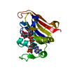 7d3zC 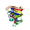 7d49C 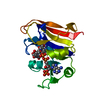 7d4xC  7d6gC 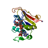 1rx2S S: Starting model for refinement C: citing same article ( |
|---|---|
| Similar structure data |


-Links
 PDBj
PDBj
- Assembly
| Deposited unit | 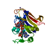
| ||||||||
|---|---|---|---|---|---|---|---|---|---|
| 1 |
| ||||||||
| Unit cell |
|
-Components
| #1: Protein | Mass: 18020.326 Da / Num. of mol.: 1 Source method: isolated from a genetically manipulated source Source: (gene. exp.) Gene: folA, tmrA, b0048, JW0047 / Plasmid: pET-sumo / Production host: |
|---|---|
| #2: Chemical | ChemComp-FOL / 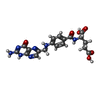  Mass: 441.397 Da / Num. of mol.: 1 / Source method: obtained synthetically / Formula: C19H19N7O6 / Feature type: SUBJECT OF INVESTIGATION Mass: 441.397 Da / Num. of mol.: 1 / Source method: obtained synthetically / Formula: C19H19N7O6 / Feature type: SUBJECT OF INVESTIGATION |
| #3: Chemical | ChemComp-NAP /   Mass: 743.405 Da / Num. of mol.: 1 / Source method: obtained synthetically / Formula: C21H28N7O17P3 Mass: 743.405 Da / Num. of mol.: 1 / Source method: obtained synthetically / Formula: C21H28N7O17P3 |
| #4: Chemical | ChemComp-MN /   Mass: 54.938 Da / Num. of mol.: 1 / Source method: isolated from a natural source / Formula: Mn Mass: 54.938 Da / Num. of mol.: 1 / Source method: isolated from a natural source / Formula: Mn |
| #5: Water | ChemComp-HOH /  Mass: 18.015 Da / Num. of mol.: 87 / Source method: isolated from a natural source / Formula: H2O Mass: 18.015 Da / Num. of mol.: 87 / Source method: isolated from a natural source / Formula: H2O |
| Has ligand of interest | Y |
-Experimental details
-Experiment
| Experiment | Method: X-RAY DIFFRACTION / Number of used crystals: 1 |
|---|
- Sample preparation
| Crystal | Density Matthews: 2.15 Å3/Da / Density % sol: 42.75 % |
|---|---|
| Crystal grow | Temperature: 291 K / Method: vapor diffusion, sitting drop / pH: 7 Details: 40mg/mL DHFR-folate-NADP+ complex, 12% (v/v) PEG 400, 100mM MnCl2, 20mM imidazole, pH7.0 |
-Data collection
| Diffraction | Mean temperature: 291 K / Serial crystal experiment: N |
|---|---|
| Diffraction source | Source: ROTATING ANODE / Type: RIGAKU MICROMAX-007 HF / Wavelength: 1.5 Å |
| Detector | Type: RIGAKU RAXIS IV++ / Detector: IMAGE PLATE / Date: Aug 10, 2014 / Details: VariMax |
| Radiation | Monochromator: CHOPPER / Protocol: SINGLE WAVELENGTH / Monochromatic (M) / Laue (L): M / Scattering type: x-ray |
| Radiation wavelength | Wavelength: 1.5 Å / Relative weight: 1 |
| Reflection | Resolution: 1.6→50 Å / Num. obs: 20795 / % possible obs: 97.7 % / Redundancy: 4.1 % / Biso Wilson estimate: 18.08 Å2 / Rmerge(I) obs: 0.097 / Χ2: 1.081 / Net I/σ(I): 13.7 |
| Reflection shell | Resolution: 1.6→1.63 Å / Redundancy: 4 % / Rmerge(I) obs: 0.446 / Mean I/σ(I) obs: 2.97 / Num. unique obs: 1017 / Χ2: 0.98 / % possible all: 94 |
- Processing
| Software |
| ||||||||||||||||||||||||||||||||||||||||||||||||
|---|---|---|---|---|---|---|---|---|---|---|---|---|---|---|---|---|---|---|---|---|---|---|---|---|---|---|---|---|---|---|---|---|---|---|---|---|---|---|---|---|---|---|---|---|---|---|---|---|---|
| Refinement | Method to determine structure: MOLECULAR REPLACEMENT Starting model: 1rx2 Resolution: 1.6→27.41 Å / SU ML: 0.16 / Cross valid method: THROUGHOUT / σ(F): 1.35 / Phase error: 18.54 / Stereochemistry target values: ML
| ||||||||||||||||||||||||||||||||||||||||||||||||
| Solvent computation | Shrinkage radii: 0.9 Å / VDW probe radii: 1.11 Å / Solvent model: FLAT BULK SOLVENT MODEL | ||||||||||||||||||||||||||||||||||||||||||||||||
| Displacement parameters | Biso max: 56.28 Å2 / Biso mean: 21.0129 Å2 / Biso min: 9.6 Å2 | ||||||||||||||||||||||||||||||||||||||||||||||||
| Refinement step | Cycle: final / Resolution: 1.6→27.41 Å
| ||||||||||||||||||||||||||||||||||||||||||||||||
| LS refinement shell | Refine-ID: X-RAY DIFFRACTION / Rfactor Rfree error: 0
|
