 Movie
Movie Controller

[English] 日本語
 Yorodumi
- PDB-4q6a: Staphylococcus aureus V31L, F98Y Mutant Dihydrofolate Reductase C... -
+ Open data

- Basic information
| Entry | Database: PDB / ID: 4q6a | ||||||
|---|---|---|---|---|---|---|---|
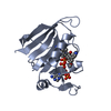
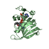
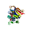
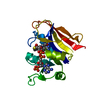
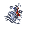
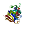
| Title | Staphylococcus aureus V31L, F98Y Mutant Dihydrofolate Reductase Complexed with NADPH | ||||||
 Components Components | Dihydrofolate reductase | ||||||
 Keywords Keywords | OXIDOREDUCTASE | ||||||
| Function / homology | Dihydrofolate Reductase, subunit A / Dihydrofolate Reductase, subunit A / 3-Layer(aba) Sandwich / Alpha Beta / ACETATE ION / NADP NICOTINAMIDE-ADENINE-DINUCLEOTIDE PHOSPHATE / :  Function and homology information Function and homology information | ||||||
| Biological species |  Staphylococcus aureus MUF168 (bacteria) Staphylococcus aureus MUF168 (bacteria) | ||||||
| Method |  X-RAY DIFFRACTION / SYNCHROTRON / MOLECULAR REPLACEMENT / Resolution: 2.099 Å X-RAY DIFFRACTION / SYNCHROTRON / MOLECULAR REPLACEMENT / Resolution: 2.099 Å | ||||||
 Authors Authors | Reeve, S.M. / Anderson, A.C. | ||||||
 Citation Citation | Journal: Proc.Natl.Acad.Sci.USA / Year: 2015 Title: Protein design algorithms predict viable resistance to an experimental antifolate. Authors: Reeve, S.M. / Gainza, P. / Frey, K.M. / Georgiev, I. / Donald, B.R. / Anderson, A.C. | ||||||
| History |
|
- Structure visualization
| Structure viewer | Molecule: MolmilJmol/JSmol |
|---|
- Downloads & links
-Download
| PDBx/mmCIF format | 4q6a.cif.gz | 52.7 KB | Display | PDBx/mmCIF format |
|---|---|---|---|---|
| PDB format | pdb4q6a.ent.gz | 36.5 KB | Display | PDB format |
| PDBx/mmJSON format | 4q6a.json.gz | Tree view | PDBx/mmJSON format | |
| Others |  Other downloads Other downloads |
-Validation report
| Arichive directory | https://data.pdbj.org/pub/pdb/validation_reports/q6/4q6aftp://data.pdbj.org/pub/pdb/validation_reports/q6/4q6a | HTTPS FTP |
|---|
-Related structure data
| Related structure data |  4q67C  3f0uS C: citing same article ( S: Starting model for refinement |
|---|---|
| Similar structure data |


-Links
 PDBj
PDBj- Assembly
| Deposited unit | 
| ||||||||
|---|---|---|---|---|---|---|---|---|---|
| 1 |
| ||||||||
| Unit cell |
|
-Components
| #1: Protein | Mass: 18419.053 Da / Num. of mol.: 1 / Mutation: V31L, F98Y Source method: isolated from a genetically manipulated source Source: (gene. exp.) Staphylococcus aureus MUF168 (bacteria)Strain: SAB1281c / Gene: dfrB, Y000_11620 / Plasmid: pET-41a(+) / Production host: |
|---|---|
| #2: Chemical | ChemComp-NAP /   Mass: 743.405 Da / Num. of mol.: 1 / Source method: obtained synthetically / Formula: C21H28N7O17P3 Mass: 743.405 Da / Num. of mol.: 1 / Source method: obtained synthetically / Formula: C21H28N7O17P3 |
| #3: Chemical | ChemComp-GOL /   Mass: 92.094 Da / Num. of mol.: 1 / Source method: obtained synthetically / Formula: C3H8O3 Mass: 92.094 Da / Num. of mol.: 1 / Source method: obtained synthetically / Formula: C3H8O3 |
| #4: Chemical | ChemComp-ACT /   Mass: 59.044 Da / Num. of mol.: 1 / Source method: obtained synthetically / Formula: C2H3O2 Mass: 59.044 Da / Num. of mol.: 1 / Source method: obtained synthetically / Formula: C2H3O2 |
| #5: Water | ChemComp-HOH /  Mass: 18.015 Da / Num. of mol.: 133 / Source method: isolated from a natural source / Formula: H2O Mass: 18.015 Da / Num. of mol.: 133 / Source method: isolated from a natural source / Formula: H2O |
-Experimental details
-Experiment
| Experiment | Method: X-RAY DIFFRACTION / Number of used crystals: 1 |
|---|
- Sample preparation
| Crystal | Density Matthews: 2.65 Å3/Da / Density % sol: 53.61 % |
|---|---|
| Crystal grow | Temperature: 277 K / Method: vapor diffusion, hanging drop / pH: 6 Details: 0.1M MES, pH 6.0, 13% PEG 10,000, 0.3M Sodium Acetate, 5% gamma-butyrolacetone, VAPOR DIFFUSION, HANGING DROP, temperature 277K |
-Data collection
| Diffraction | Mean temperature: 100 K |
|---|---|
| Diffraction source | Source: SYNCHROTRON / Site: NSLS  / Beamline: X4A / Wavelength: 0.97915 Å / Beamline: X4A / Wavelength: 0.97915 Å |
| Detector | Type: MAR CCD 165 mm / Detector: CCD / Date: Sep 20, 2013 |
| Radiation | Monochromator: KOHZU double crystal monochromator / Protocol: SINGLE WAVELENGTH / Monochromatic (M) / Laue (L): M / Scattering type: x-ray |
| Radiation wavelength | Wavelength: 0.97915 Å / Relative weight: 1 |
| Reflection | Resolution: 2.099→34.351 Å / Num. all: 12235 / Num. obs: 11649 / % possible obs: 99.87 % / Observed criterion σ(F): 0 / Observed criterion σ(I): 0 / Redundancy: 13.6 % / Biso Wilson estimate: 19.57 Å2 / Rmerge(I) obs: 0.094 |
| Reflection shell | Resolution: 2.1→2.14 Å / Redundancy: 13.9 % / Rmerge(I) obs: 0.354 / Mean I/σ(I) obs: 20 / % possible all: 0.993 |
- Processing
| Software |
| ||||||||||||||||||||||||||||||
|---|---|---|---|---|---|---|---|---|---|---|---|---|---|---|---|---|---|---|---|---|---|---|---|---|---|---|---|---|---|---|---|
| Refinement | Method to determine structure: MOLECULAR REPLACEMENT Starting model: pdb entry 3F0U Resolution: 2.099→34.351 Å / SU ML: 0.16 / σ(F): 1.34 / Phase error: 19.85 / Stereochemistry target values: ML
| ||||||||||||||||||||||||||||||
| Solvent computation | Shrinkage radii: 0.9 Å / VDW probe radii: 1.11 Å / Solvent model: FLAT BULK SOLVENT MODEL | ||||||||||||||||||||||||||||||
| Refinement step | Cycle: LAST / Resolution: 2.099→34.351 Å
| ||||||||||||||||||||||||||||||
| Refine LS restraints |
| ||||||||||||||||||||||||||||||
| LS refinement shell | Refine-ID: X-RAY DIFFRACTION / % reflection obs: 100 %
|
