 Movie
Movie Controller

[English] 日本語
 Yorodumi
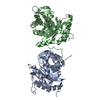
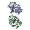
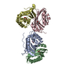
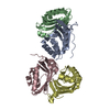
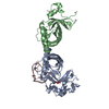
- PDB-7d2p: Crystal structure of MazE-MazF (Form-II) from Deinococcus radiodurans -
+ Open data

- Basic information
| Entry | Database: PDB / ID: 7d2p | ||||||
|---|---|---|---|---|---|---|---|
| Title | Crystal structure of MazE-MazF (Form-II) from Deinococcus radiodurans | ||||||
 Components Components |
| ||||||
 Keywords Keywords | TOXIN / MazEF toxin-antitoxin system / Antitoxin / endonuclease | ||||||
| Function / homology |  Function and homology information Function and homology informationtoxin sequestering activity / toxin-antitoxin complex / rRNA catabolic process / Hydrolases; Acting on ester bonds; Endoribonucleases producing 3'-phosphomonoesters / mRNA catabolic process / RNA endonuclease activity / hydrolase activity / regulation of DNA-templated transcription / DNA binding Similarity search - Function | ||||||
| Biological species |  Deinococcus radiodurans (radioresistant) Deinococcus radiodurans (radioresistant) | ||||||
| Method |  X-RAY DIFFRACTION / SYNCHROTRON / MOLECULAR REPLACEMENT / Resolution: 2.07 Å X-RAY DIFFRACTION / SYNCHROTRON / MOLECULAR REPLACEMENT / Resolution: 2.07 Å | ||||||
 Authors Authors | Dhanasingh, I. / Lee, S.H. | ||||||
 Citation Citation | Journal: J.Microbiol / Year: 2021 Title: Functional and structural characterization of Deinococcus radiodurans R1 MazEF toxin-antitoxin system, Dr0416-Dr0417. Authors: Dhanasingh, I. / Choi, E. / Lee, J. / Lee, S.H. / Hwang, J. | ||||||
| History |
|
- Structure visualization
| Structure viewer | Molecule: MolmilJmol/JSmol |
|---|
- Downloads & links
-Download
| PDBx/mmCIF format | 7d2p.cif.gz | 110.6 KB | Display | PDBx/mmCIF format |
|---|---|---|---|---|
| PDB format | pdb7d2p.ent.gz | 83.1 KB | Display | PDB format |
| PDBx/mmJSON format | 7d2p.json.gz | Tree view | PDBx/mmJSON format | |
| Others |  Other downloads Other downloads |
-Validation report
| Arichive directory | https://data.pdbj.org/pub/pdb/validation_reports/d2/7d2pftp://data.pdbj.org/pub/pdb/validation_reports/d2/7d2p | HTTPS FTP |
|---|
-Related structure data
| Related structure data |  7d28SC  7d2mC  7d2nC  7d2qC S: Starting model for refinement C: citing same article ( |
|---|---|
| Similar structure data |

-Links
 PDBj
PDBj- Assembly
| Deposited unit | 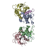
| ||||||||
|---|---|---|---|---|---|---|---|---|---|
| 1 |
| ||||||||
| Unit cell |
|
-Components
| #1: Protein | Mass: 12465.216 Da / Num. of mol.: 4 Source method: isolated from a genetically manipulated source Source: (gene. exp.) Deinococcus radiodurans (radioresistant)Gene: mazF, HAV23_04965, HAV35_11860 / Production host: References: UniProt: A0A6G9BVQ8, UniProt: Q9RX98*PLUS, Hydrolases; Acting on ester bonds; Endoribonucleases producing 3'-phosphomonoesters #2: Protein | Mass: 8918.065 Da / Num. of mol.: 2 Source method: isolated from a genetically manipulated source Source: (gene. exp.) Deinococcus radiodurans (radioresistant)Gene: HAV23_04960, HAV35_11865 / Production host: #3: Water | ChemComp-HOH / |  Mass: 18.015 Da / Num. of mol.: 237 / Source method: isolated from a natural source / Formula: H2O Mass: 18.015 Da / Num. of mol.: 237 / Source method: isolated from a natural source / Formula: H2O |
|---|
-Experimental details
-Experiment
| Experiment | Method: X-RAY DIFFRACTION / Number of used crystals: 1 |
|---|
- Sample preparation
| Crystal | Density Matthews: 1.59 Å3/Da / Density % sol: 22.72 % |
|---|---|
| Crystal grow | Temperature: 293 K / Method: vapor diffusion, hanging drop / Details: 0.1 M succinic acid pH 7.0 and 12% w/v PEG 3,350 |
-Data collection
| Diffraction | Mean temperature: 193 K / Serial crystal experiment: N | |||||||||||||||||||||||||||||||||||||||||||||||||||||||||||||||||||||||||||||||||||||||||||||||||||||||||||||||||||||||||||||||||||||||||||||||||||||||||||||||||||||||||||||||||||||||||||||
|---|---|---|---|---|---|---|---|---|---|---|---|---|---|---|---|---|---|---|---|---|---|---|---|---|---|---|---|---|---|---|---|---|---|---|---|---|---|---|---|---|---|---|---|---|---|---|---|---|---|---|---|---|---|---|---|---|---|---|---|---|---|---|---|---|---|---|---|---|---|---|---|---|---|---|---|---|---|---|---|---|---|---|---|---|---|---|---|---|---|---|---|---|---|---|---|---|---|---|---|---|---|---|---|---|---|---|---|---|---|---|---|---|---|---|---|---|---|---|---|---|---|---|---|---|---|---|---|---|---|---|---|---|---|---|---|---|---|---|---|---|---|---|---|---|---|---|---|---|---|---|---|---|---|---|---|---|---|---|---|---|---|---|---|---|---|---|---|---|---|---|---|---|---|---|---|---|---|---|---|---|---|---|---|---|---|---|---|---|---|---|
| Diffraction source | Source: SYNCHROTRON / Site: PAL/PLS  / Beamline: 7A (6B, 6C1) / Wavelength: 0.97935 Å / Beamline: 7A (6B, 6C1) / Wavelength: 0.97935 Å | |||||||||||||||||||||||||||||||||||||||||||||||||||||||||||||||||||||||||||||||||||||||||||||||||||||||||||||||||||||||||||||||||||||||||||||||||||||||||||||||||||||||||||||||||||||||||||||
| Detector | Type: ADSC QUANTUM 270 / Detector: CCD / Date: Oct 2, 2017 | |||||||||||||||||||||||||||||||||||||||||||||||||||||||||||||||||||||||||||||||||||||||||||||||||||||||||||||||||||||||||||||||||||||||||||||||||||||||||||||||||||||||||||||||||||||||||||||
| Radiation | Protocol: SINGLE WAVELENGTH / Monochromatic (M) / Laue (L): M / Scattering type: x-ray | |||||||||||||||||||||||||||||||||||||||||||||||||||||||||||||||||||||||||||||||||||||||||||||||||||||||||||||||||||||||||||||||||||||||||||||||||||||||||||||||||||||||||||||||||||||||||||||
| Radiation wavelength | Wavelength: 0.97935 Å / Relative weight: 1 | |||||||||||||||||||||||||||||||||||||||||||||||||||||||||||||||||||||||||||||||||||||||||||||||||||||||||||||||||||||||||||||||||||||||||||||||||||||||||||||||||||||||||||||||||||||||||||||
| Reflection | Resolution: 2→50 Å / Num. obs: 28134 / % possible obs: 97.2 % / Redundancy: 3.7 % / Rmerge(I) obs: 0.063 / Rpim(I) all: 0.038 / Rrim(I) all: 0.074 / Χ2: 0.791 / Net I/σ(I): 8.4 / Num. measured all: 104692 | |||||||||||||||||||||||||||||||||||||||||||||||||||||||||||||||||||||||||||||||||||||||||||||||||||||||||||||||||||||||||||||||||||||||||||||||||||||||||||||||||||||||||||||||||||||||||||||
| Reflection shell | Diffraction-ID: 1
|
- Processing
| Software |
| ||||||||||||||||||||||||||||||||||||||||||||||||||||||||||||
|---|---|---|---|---|---|---|---|---|---|---|---|---|---|---|---|---|---|---|---|---|---|---|---|---|---|---|---|---|---|---|---|---|---|---|---|---|---|---|---|---|---|---|---|---|---|---|---|---|---|---|---|---|---|---|---|---|---|---|---|---|---|
| Refinement | Method to determine structure: MOLECULAR REPLACEMENT Starting model: 7D28 Resolution: 2.07→29.4 Å / Cor.coef. Fo:Fc: 0.955 / Cor.coef. Fo:Fc free: 0.918 / SU B: 5.087 / SU ML: 0.135 / Cross valid method: THROUGHOUT / σ(F): 0 / ESU R: 0.291 / ESU R Free: 0.213 / Stereochemistry target values: MAXIMUM LIKELIHOOD Details: HYDROGENS HAVE BEEN ADDED IN THE RIDING POSITIONS U VALUES : REFINED INDIVIDUALLY
| ||||||||||||||||||||||||||||||||||||||||||||||||||||||||||||
| Solvent computation | Ion probe radii: 0.8 Å / Shrinkage radii: 0.8 Å / VDW probe radii: 1.2 Å / Solvent model: MASK | ||||||||||||||||||||||||||||||||||||||||||||||||||||||||||||
| Displacement parameters | Biso max: 81.74 Å2 / Biso mean: 25.914 Å2 / Biso min: 11.62 Å2
| ||||||||||||||||||||||||||||||||||||||||||||||||||||||||||||
| Refinement step | Cycle: final / Resolution: 2.07→29.4 Å
| ||||||||||||||||||||||||||||||||||||||||||||||||||||||||||||
| Refine LS restraints |
| ||||||||||||||||||||||||||||||||||||||||||||||||||||||||||||
| LS refinement shell | Resolution: 2.074→2.127 Å / Rfactor Rfree error: 0 / Total num. of bins used: 20
|
