 Movie
Movie Controller


+ Open data

- Basic information
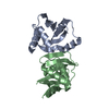
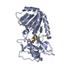
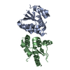
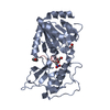
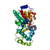
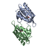
| Entry | Database: PDB / ID: 7d0e | |||||||||
|---|---|---|---|---|---|---|---|---|---|---|
| Title | Crystal structure of FIP200 Claw/p-CCPG1 FIR2 | |||||||||
 Components Components |
| |||||||||
 Keywords Keywords | SIGNALING PROTEIN/PROTEIN BINDING / autophagy / FIP200 / CCPG1 / SIGNALING PROTEIN / SIGNALING PROTEIN-PROTEIN BINDING complex | |||||||||
| Function / homology |  Function and homology information Function and homology informationAtg1/ULK1 kinase complex / ribophagy / glycophagy / phagophore assembly site membrane / pexophagy / autophagy of mitochondrion / piecemeal microautophagy of the nucleus / phagophore assembly site / reticulophagy / Macroautophagy ...Atg1/ULK1 kinase complex / ribophagy / glycophagy / phagophore assembly site membrane / pexophagy / autophagy of mitochondrion / piecemeal microautophagy of the nucleus / phagophore assembly site / reticulophagy / Macroautophagy / autophagosome membrane / autophagosome assembly / positive regulation of cell size / positive regulation of cell cycle / extrinsic apoptotic signaling pathway / protein-membrane adaptor activity / positive regulation of autophagy / negative regulation of extrinsic apoptotic signaling pathway / positive regulation of JNK cascade / liver development / autophagy / heart development / nuclear membrane / defense response to virus / molecular adaptor activity / lysosome / negative regulation of cell population proliferation / innate immune response / positive regulation of cell population proliferation / endoplasmic reticulum membrane / protein kinase binding / positive regulation of transcription by RNA polymerase II / membrane / cytosol Similarity search - Function | |||||||||
| Biological species |  Homo sapiens (human) Homo sapiens (human) | |||||||||
| Method |  X-RAY DIFFRACTION / SYNCHROTRON / MOLECULAR REPLACEMENT / Resolution: 1.4 Å X-RAY DIFFRACTION / SYNCHROTRON / MOLECULAR REPLACEMENT / Resolution: 1.4 Å | |||||||||
 Authors Authors | Zhou, Z.X. / Pan, L.F. | |||||||||
| Funding support |  China, 2items China, 2items
| |||||||||
 Citation Citation | Journal: Nat Commun / Year: 2021 Title: Phosphorylation regulates the binding of autophagy receptors to FIP200 Claw domain for selective autophagy initiation. Authors: Zhou, Z. / Liu, J. / Fu, T. / Wu, P. / Peng, C. / Gong, X. / Wang, Y. / Zhang, M. / Li, Y. / Wang, Y. / Xu, X. / Li, M. / Pan, L. | |||||||||
| History |
|
- Structure visualization
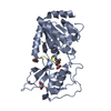
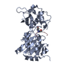
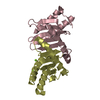
| Structure viewer | Molecule: MolmilJmol/JSmol |
|---|
- Downloads & links
-Download
| PDBx/mmCIF format | 7d0e.cif.gz | 69.3 KB | Display | PDBx/mmCIF format |
|---|---|---|---|---|
| PDB format | pdb7d0e.ent.gz | 47 KB | Display | PDB format |
| PDBx/mmJSON format | 7d0e.json.gz | Tree view | PDBx/mmJSON format | |
| Others |  Other downloads Other downloads |
-Validation report
| Summary document | 7d0e_validation.pdf.gz | 847.9 KB | Display | wwPDB validaton report |
|---|---|---|---|---|
| Full document | 7d0e_full_validation.pdf.gz | 849.1 KB | Display | |
| Data in XML | 7d0e_validation.xml.gz | 8.5 KB | Display | |
| Data in CIF | 7d0e_validation.cif.gz | 11.2 KB | Display | |
| Arichive directory | https://data.pdbj.org/pub/pdb/validation_reports/d0/7d0eftp://data.pdbj.org/pub/pdb/validation_reports/d0/7d0e | HTTPS FTP |
-Related structure data
| Related structure data |  7czgC  7czmC  6dceS S: Starting model for refinement C: citing same article ( |
|---|---|
| Similar structure data |
-Links
 PDBj
PDBj
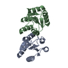
- Assembly
| Deposited unit | 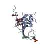
| ||||||||||||
|---|---|---|---|---|---|---|---|---|---|---|---|---|---|
| 1 | 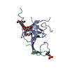
| ||||||||||||
| Unit cell |
| ||||||||||||
| Components on special symmetry positions |
|
-Components
| #1: Protein | Mass: 12039.970 Da / Num. of mol.: 1 Source method: isolated from a genetically manipulated source Source: (gene. exp.) Homo sapiens (human) / Gene: RB1CC1, KIAA0203, RBICC / Production host:  |
|---|---|
| #2: Protein/peptide | Mass: 1496.487 Da / Num. of mol.: 1 Source method: isolated from a genetically manipulated source Source: (gene. exp.) Homo sapiens (human) / Gene: CCPG1 / Production host: |
| #3: Chemical | ChemComp-GO9 /   Mass: 312.357 Da / Num. of mol.: 1 / Source method: isolated from a natural source / Formula: C13H28O8 Mass: 312.357 Da / Num. of mol.: 1 / Source method: isolated from a natural source / Formula: C13H28O8 |
| #4: Chemical | ChemComp-PEG /   Mass: 106.120 Da / Num. of mol.: 1 / Source method: obtained synthetically / Formula: C4H10O3 Mass: 106.120 Da / Num. of mol.: 1 / Source method: obtained synthetically / Formula: C4H10O3 |
| #5: Water | ChemComp-HOH /  Mass: 18.015 Da / Num. of mol.: 95 / Source method: isolated from a natural source / Formula: H2O Mass: 18.015 Da / Num. of mol.: 95 / Source method: isolated from a natural source / Formula: H2O |
| Has ligand of interest | Y |
| Has protein modification | Y |
-Experimental details
-Experiment
| Experiment | Method: X-RAY DIFFRACTION / Number of used crystals: 1 |
|---|
- Sample preparation
| Crystal | Density Matthews: 2.1 Å3/Da / Density % sol: 41.57 % |
|---|---|
| Crystal grow | Temperature: 289 K / Method: vapor diffusion, sitting drop / Details: KCl, Pentaerythritol ethoxylate (15/4 EO/OH), MES |
-Data collection
| Diffraction | Mean temperature: 100 K / Serial crystal experiment: N |
|---|---|
| Diffraction source | Source: SYNCHROTRON / Site: SSRF / Beamline: BL19U1 / Wavelength: 0.97876 Å |
| Detector | Type: DECTRIS PILATUS3 S 6M / Detector: PIXEL / Date: Apr 12, 2019 |
| Radiation | Protocol: SINGLE WAVELENGTH / Monochromatic (M) / Laue (L): M / Scattering type: x-ray |
| Radiation wavelength | Wavelength: 0.97876 Å / Relative weight: 1 |
| Reflection | Resolution: 1.4→50 Å / Num. obs: 23548 / % possible obs: 99.8 % / Redundancy: 25.1 % / Biso Wilson estimate: 12.28 Å2 / Rmerge(I) obs: 0.037 / Net I/σ(I): 98.9 |
| Reflection shell | Resolution: 1.4→1.42 Å / Num. unique obs: 1143 / Rpim(I) all: 0.174 |
- Processing
| Software |
| ||||||||||||||||||||||||
|---|---|---|---|---|---|---|---|---|---|---|---|---|---|---|---|---|---|---|---|---|---|---|---|---|---|
| Refinement | Method to determine structure: MOLECULAR REPLACEMENT Starting model: 6DCE Resolution: 1.4→25.07 Å / Cross valid method: FREE R-VALUE Stereochemistry target values: GeoStd + Monomer Library + CDL v1.2
| ||||||||||||||||||||||||
| Displacement parameters | Biso mean: 24.49 Å2 | ||||||||||||||||||||||||
| Refinement step | Cycle: LAST / Resolution: 1.4→25.07 Å
| ||||||||||||||||||||||||
| Refine LS restraints |
|
