 Movie
Movie Controller


+ Open data

- Basic information
| Entry | Database: PDB / ID: 7czm | |||||||||
|---|---|---|---|---|---|---|---|---|---|---|
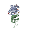
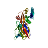
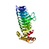
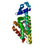
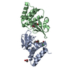
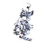
| Title | Crystal structure of FIP200 Claw/p-OPtineurin LIR complex | |||||||||
 Components Components |
| |||||||||
 Keywords Keywords | SIGNALING PROTEIN/PROTEIN BINDING / autophagy / FIP200 / Optineurin / SIGNALING PROTEIN / SIGNALING PROTEIN-PROTEIN BINDING complex | |||||||||
| Function / homology |  Function and homology information Function and homology informationregulation of protein lipidation / type 2 mitophagy / Atg1/ULK1 kinase complex / cell death / glycophagy / negative regulation of receptor recycling / protein localization to Golgi apparatus / Golgi ribbon formation / positive regulation of xenophagy / ribophagy ...regulation of protein lipidation / type 2 mitophagy / Atg1/ULK1 kinase complex / cell death / glycophagy / negative regulation of receptor recycling / protein localization to Golgi apparatus / Golgi ribbon formation / positive regulation of xenophagy / ribophagy / phagophore assembly site membrane / autophagy of mitochondrion / piecemeal microautophagy of the nucleus / pexophagy / Golgi to plasma membrane protein transport / phagophore assembly site / TBC/RABGAPs / regulation of canonical NF-kappaB signal transduction / reticulophagy / TNFR1-induced proapoptotic signaling / K63-linked polyubiquitin modification-dependent protein binding / Macroautophagy / Golgi organization / autophagosome membrane / cellular response to unfolded protein / polyubiquitin modification-dependent protein binding / autophagosome assembly / protein-membrane adaptor activity / positive regulation of autophagy / autophagosome / Regulation of TBK1, IKKε (IKBKE)-mediated activation of IRF3, IRF7 / Regulation of TBK1, IKKε-mediated activation of IRF3, IRF7 upon TLR3 ligation / TICAM1-dependent activation of IRF3/IRF7 / negative regulation of canonical NF-kappaB signal transduction / Activation of IRF3, IRF7 mediated by TBK1, IKKε (IKBKE) / PINK1-PRKN Mediated Mitophagy / macroautophagy / TNFR1-induced NF-kappa-B signaling pathway / trans-Golgi network / Regulation of TNFR1 signaling / autophagy / recycling endosome membrane / Regulation of PLK1 Activity at G2/M Transition / defense response to Gram-negative bacterium / molecular adaptor activity / protein-macromolecule adaptor activity / lysosome / negative regulation of cell population proliferation / Golgi membrane / innate immune response / protein kinase binding / endoplasmic reticulum membrane / perinuclear region of cytoplasm / Golgi apparatus / signal transduction / DNA-templated transcription / zinc ion binding / nucleoplasm / identical protein binding / nucleus / cytosol / cytoplasm Similarity search - Function | |||||||||
| Biological species |  Homo sapiens (human) Homo sapiens (human) | |||||||||
| Method |  X-RAY DIFFRACTION / SYNCHROTRON / MOLECULAR REPLACEMENT / Resolution: 2 Å X-RAY DIFFRACTION / SYNCHROTRON / MOLECULAR REPLACEMENT / Resolution: 2 Å | |||||||||
 Authors Authors | Zhou, Z.X. / Pan, L.F. | |||||||||
| Funding support |  China, 2items China, 2items
| |||||||||
 Citation Citation | Journal: Nat Commun / Year: 2021 Title: Phosphorylation regulates the binding of autophagy receptors to FIP200 Claw domain for selective autophagy initiation. Authors: Zhou, Z. / Liu, J. / Fu, T. / Wu, P. / Peng, C. / Gong, X. / Wang, Y. / Zhang, M. / Li, Y. / Wang, Y. / Xu, X. / Li, M. / Pan, L. | |||||||||
| History |
|
- Structure visualization
| Structure viewer | Molecule: MolmilJmol/JSmol |
|---|
- Downloads & links
-Download
| PDBx/mmCIF format | 7czm.cif.gz | 110.1 KB | Display | PDBx/mmCIF format |
|---|---|---|---|---|
| PDB format | pdb7czm.ent.gz | 79.9 KB | Display | PDB format |
| PDBx/mmJSON format | 7czm.json.gz | Tree view | PDBx/mmJSON format | |
| Others |  Other downloads Other downloads |
-Validation report
| Arichive directory | https://data.pdbj.org/pub/pdb/validation_reports/cz/7czmftp://data.pdbj.org/pub/pdb/validation_reports/cz/7czm | HTTPS FTP |
|---|
-Related structure data
| Related structure data |  7czgC 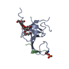 7d0eC  6dceS S: Starting model for refinement C: citing same article ( |
|---|---|
| Similar structure data |


-Links
 PDBj
PDBj


- Assembly
| Deposited unit | 
| ||||||||||||
|---|---|---|---|---|---|---|---|---|---|---|---|---|---|
| 1 |
| ||||||||||||
| Unit cell |
| ||||||||||||
| Components on special symmetry positions |
|
-Components
| #1: Protein | Mass: 12403.336 Da / Num. of mol.: 2 Source method: isolated from a genetically manipulated source Source: (gene. exp.) Homo sapiens (human) / Gene: RB1CC1, KIAA0203, RBICC / Production host:  #2: Protein/peptide | Mass: 1580.587 Da / Num. of mol.: 2 Source method: isolated from a genetically manipulated source Source: (gene. exp.) Homo sapiens (human) / Gene: OPTN, FIP2, GLC1E, HIP7, HYPL, NRP / Production host: #3: Chemical |   Mass: 92.094 Da / Num. of mol.: 2 / Source method: obtained synthetically / Formula: C3H8O3 Mass: 92.094 Da / Num. of mol.: 2 / Source method: obtained synthetically / Formula: C3H8O3#4: Chemical | ChemComp-CL /   Mass: 35.453 Da / Num. of mol.: 4 / Source method: obtained synthetically / Formula: Cl Mass: 35.453 Da / Num. of mol.: 4 / Source method: obtained synthetically / Formula: Cl#5: Water | ChemComp-HOH / |  Mass: 18.015 Da / Num. of mol.: 85 / Source method: isolated from a natural source / Formula: H2O Mass: 18.015 Da / Num. of mol.: 85 / Source method: isolated from a natural source / Formula: H2OHas ligand of interest | Y | Has protein modification | Y | |
|---|
-Experimental details
-Experiment
| Experiment | Method: X-RAY DIFFRACTION / Number of used crystals: 1 |
|---|
- Sample preparation
| Crystal | Density Matthews: 2.56 Å3/Da / Density % sol: 51.88 % |
|---|---|
| Crystal grow | Temperature: 289 K / Method: vapor diffusion, sitting drop / Details: Potassium iodide, MES pH 6.5, PEG 4000 |
-Data collection
| Diffraction | Mean temperature: 100 K / Serial crystal experiment: N |
|---|---|
| Diffraction source | Source: SYNCHROTRON / Site: SSRF / Beamline: BL17U1 / Wavelength: 0.97918 Å |
| Detector | Type: DECTRIS PILATUS3 S 6M / Detector: PIXEL / Date: May 3, 2020 |
| Radiation | Protocol: SINGLE WAVELENGTH / Monochromatic (M) / Laue (L): M / Scattering type: x-ray |
| Radiation wavelength | Wavelength: 0.97918 Å / Relative weight: 1 |
| Reflection | Resolution: 2→35.32 Å / Num. obs: 18560 / % possible obs: 99.6 % / Redundancy: 6.6 % / Biso Wilson estimate: 41.48 Å2 / Rmerge(I) obs: 0.034 / Net I/σ(I): 31.6 |
| Reflection shell | Resolution: 2→2.07 Å / Rmerge(I) obs: 0.254 / Num. unique obs: 1836 |
- Processing
| Software |
| ||||||||||||||||||||||||
|---|---|---|---|---|---|---|---|---|---|---|---|---|---|---|---|---|---|---|---|---|---|---|---|---|---|
| Refinement | Method to determine structure: MOLECULAR REPLACEMENT Starting model: 6DCE Resolution: 2→34.96 Å / Cross valid method: FREE R-VALUE Stereochemistry target values: GeoStd + Monomer Library + CDL v1.2
| ||||||||||||||||||||||||
| Displacement parameters | Biso mean: 56.42 Å2 | ||||||||||||||||||||||||
| Refinement step | Cycle: LAST / Resolution: 2→34.96 Å
| ||||||||||||||||||||||||
| Refine LS restraints |
|
