 Movie
Movie Controller


+ Open data

- Basic information
| Entry | Database: PDB / ID: 7czd | ||||||
|---|---|---|---|---|---|---|---|
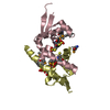
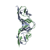
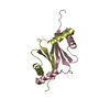
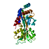
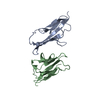
| Title | Crystal structure of PD-L1 in complex with a VHH | ||||||
 Components Components |
| ||||||
 Keywords Keywords | IMMUNE SYSTEM / antibody / Complex / VHH | ||||||
| Function / homology |  Function and homology information Function and homology informationnegative regulation of tumor necrosis factor superfamily cytokine production / positive regulation of activated CD8-positive, alpha-beta T cell apoptotic process / negative regulation of CD8-positive, alpha-beta T cell activation / TRIF-dependent toll-like receptor signaling pathway / negative regulation of T cell mediated immune response to tumor cell / negative regulation of CD4-positive, alpha-beta T cell proliferation / STAT3 nuclear events downstream of ALK signaling / negative regulation of interleukin-10 production / negative regulation of T cell activation / negative regulation of activated T cell proliferation ...negative regulation of tumor necrosis factor superfamily cytokine production / positive regulation of activated CD8-positive, alpha-beta T cell apoptotic process / negative regulation of CD8-positive, alpha-beta T cell activation / TRIF-dependent toll-like receptor signaling pathway / negative regulation of T cell mediated immune response to tumor cell / negative regulation of CD4-positive, alpha-beta T cell proliferation / STAT3 nuclear events downstream of ALK signaling / negative regulation of interleukin-10 production / negative regulation of T cell activation / negative regulation of activated T cell proliferation / negative regulation of type II interferon production / positive regulation of interleukin-10 production / Co-inhibition by PD-1 / negative regulation of T cell receptor signaling pathway / negative regulation of T cell proliferation / T cell costimulation / response to cytokine / positive regulation of T cell proliferation / recycling endosome membrane / actin cytoskeleton / cellular response to lipopolysaccharide / early endosome membrane / adaptive immune response / transcription coactivator activity / cell surface receptor signaling pathway / immune response / receptor ligand activity / external side of plasma membrane / signal transduction / extracellular exosome / nucleoplasm / plasma membrane Similarity search - Function | ||||||
| Biological species |   Homo sapiens (human) Homo sapiens (human) | ||||||
| Method |  X-RAY DIFFRACTION / SYNCHROTRON / MOLECULAR REPLACEMENT / Resolution: 1.64 Å X-RAY DIFFRACTION / SYNCHROTRON / MOLECULAR REPLACEMENT / Resolution: 1.64 Å | ||||||
 Authors Authors | Wang, C. | ||||||
 Citation Citation | Journal: J Immunother Cancer / Year: 2021 Title: Generation of a safe and efficacious llama single-domain antibody fragment (vHH) targeting the membrane-proximal region of 4-1BB for engineering therapeutic bispecific antibodies for cancer. Authors: Zhai, T. / Wang, C. / Xu, Y. / Huang, W. / Yuan, Z. / Wang, T. / Dai, S. / Peng, S. / Pang, T. / Jiang, W. / Huang, Y. / Zou, Y. / Xu, Y. / Sun, J. / Gong, X. / Zhang, J. / Tsun, A. / Li, B. / Miao, X. | ||||||
| History |
|
- Structure visualization
| Structure viewer | Molecule: MolmilJmol/JSmol |
|---|
- Downloads & links
-Download
| PDBx/mmCIF format | 7czd.cif.gz | 119.7 KB | Display | PDBx/mmCIF format |
|---|---|---|---|---|
| PDB format | pdb7czd.ent.gz | 91.1 KB | Display | PDB format |
| PDBx/mmJSON format | 7czd.json.gz | Tree view | PDBx/mmJSON format | |
| Others |  Other downloads Other downloads |
-Validation report
| Arichive directory | https://data.pdbj.org/pub/pdb/validation_reports/cz/7czdftp://data.pdbj.org/pub/pdb/validation_reports/cz/7czd | HTTPS FTP |
|---|
-Related structure data
| Related structure data |  7d4bC  5jdsS  5m2jS S: Starting model for refinement C: citing same article ( |
|---|---|
| Similar structure data |


-Links
 PDBj
PDBj


- Assembly
| Deposited unit | 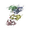
| ||||||||
|---|---|---|---|---|---|---|---|---|---|
| 1 | 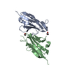
| ||||||||
| 2 | 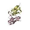
| ||||||||
| Unit cell |
|
-Components
| #1: Antibody | Mass: 12657.917 Da / Num. of mol.: 2 Source method: isolated from a genetically manipulated source Source: (gene. exp.) Homo sapiens (human)#2: Protein | Mass: 13454.492 Da / Num. of mol.: 2 Source method: isolated from a genetically manipulated source Source: (gene. exp.) Homo sapiens (human) / Gene: CD274, B7H1, PDCD1L1, PDCD1LG1, PDL1 / Production host:  #3: Chemical | ChemComp-EDO /   Mass: 62.068 Da / Num. of mol.: 6 / Source method: obtained synthetically / Formula: C2H6O2 Mass: 62.068 Da / Num. of mol.: 6 / Source method: obtained synthetically / Formula: C2H6O2#4: Chemical | ChemComp-PEG / |   Mass: 106.120 Da / Num. of mol.: 1 / Source method: obtained synthetically / Formula: C4H10O3 Mass: 106.120 Da / Num. of mol.: 1 / Source method: obtained synthetically / Formula: C4H10O3#5: Water | ChemComp-HOH / |  Mass: 18.015 Da / Num. of mol.: 586 / Source method: isolated from a natural source / Formula: H2O Mass: 18.015 Da / Num. of mol.: 586 / Source method: isolated from a natural source / Formula: H2OHas ligand of interest | N | Has protein modification | Y | |
|---|
-Experimental details
-Experiment
| Experiment | Method: X-RAY DIFFRACTION / Number of used crystals: 1 |
|---|
- Sample preparation
| Crystal | Density Matthews: 2.19 Å3/Da / Density % sol: 43.91 % |
|---|---|
| Crystal grow | Temperature: 291 K / Method: vapor diffusion, sitting drop Details: 0.2 M ammonium sulfate, 0.1 M Bis-Tris pH 5.5, 25% w/v PEG 3350 |
-Data collection
| Diffraction | Mean temperature: 100 K / Serial crystal experiment: N | |||||||||||||||
|---|---|---|---|---|---|---|---|---|---|---|---|---|---|---|---|---|
| Diffraction source | Source: SYNCHROTRON / Site: SSRF  / Beamline: BL19U1 / Wavelength: 0.978 Å / Beamline: BL19U1 / Wavelength: 0.978 Å | |||||||||||||||
| Detector | Type: DECTRIS PILATUS3 6M / Detector: PIXEL / Date: Oct 25, 2019 | |||||||||||||||
| Radiation | Protocol: SINGLE WAVELENGTH / Monochromatic (M) / Laue (L): M / Scattering type: x-ray | |||||||||||||||
| Radiation wavelength | Wavelength: 0.978 Å / Relative weight: 1 | |||||||||||||||
| Reflection twin |
| |||||||||||||||
| Reflection | Resolution: 1.64→39.65 Å / Num. obs: 51992 / % possible obs: 94.6 % / Redundancy: 3.5 % / CC1/2: 0.999 / Rmerge(I) obs: 0.041 / Rpim(I) all: 0.031 / Net I/σ(I): 15 | |||||||||||||||
| Reflection shell | Resolution: 1.64→1.67 Å / Rmerge(I) obs: 0.441 / Num. unique obs: 2495 / CC1/2: 0.796 / % possible all: 92.6 |
- Processing
| Software |
| ||||||||||||||||||||||||||||||||||||||||||||||||||||||||||||
|---|---|---|---|---|---|---|---|---|---|---|---|---|---|---|---|---|---|---|---|---|---|---|---|---|---|---|---|---|---|---|---|---|---|---|---|---|---|---|---|---|---|---|---|---|---|---|---|---|---|---|---|---|---|---|---|---|---|---|---|---|---|
| Refinement | Method to determine structure: MOLECULAR REPLACEMENT Starting model: 5JDS, 5M2J Resolution: 1.64→39.65 Å / Cor.coef. Fo:Fc: 0.971 / Cor.coef. Fo:Fc free: 0.952 / SU B: 1.496 / SU ML: 0.055 / Cross valid method: THROUGHOUT / σ(F): 0 / ESU R: 0.02 / ESU R Free: 0.02 / Stereochemistry target values: MAXIMUM LIKELIHOOD Details: HYDROGENS HAVE BEEN ADDED IN THE RIDING POSITIONS U VALUES : REFINED INDIVIDUALLY
| ||||||||||||||||||||||||||||||||||||||||||||||||||||||||||||
| Solvent computation | Ion probe radii: 0.8 Å / Shrinkage radii: 0.8 Å / VDW probe radii: 1.2 Å / Solvent model: MASK | ||||||||||||||||||||||||||||||||||||||||||||||||||||||||||||
| Displacement parameters | Biso max: 58.04 Å2 / Biso mean: 19.532 Å2 / Biso min: 11.92 Å2
| ||||||||||||||||||||||||||||||||||||||||||||||||||||||||||||
| Refinement step | Cycle: final / Resolution: 1.64→39.65 Å
| ||||||||||||||||||||||||||||||||||||||||||||||||||||||||||||
| Refine LS restraints |
| ||||||||||||||||||||||||||||||||||||||||||||||||||||||||||||
| LS refinement shell | Resolution: 1.64→1.682 Å / Rfactor Rfree error: 0 / Total num. of bins used: 20
|
