[English] 日本語
 Yorodumi
- PDB-1qa9: Structure of a Heterophilic Adhesion Complex Between the Human CD... -
+ Open data
- Basic information
| Entry | Database: PDB / ID: 1qa9 | ||||||
|---|---|---|---|---|---|---|---|
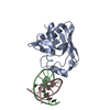
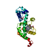
| Title | Structure of a Heterophilic Adhesion Complex Between the Human CD2 and CD58(LFA-3) Counter-Receptors | ||||||
 Components Components |
| ||||||
 Keywords Keywords | IMMUNE SYSTEM / CELL ADHESION / IG-LIKE DOMAIN / CD2 / CD58 | ||||||
| Function / homology |  Function and homology information Function and homology informationpositive regulation of myeloid dendritic cell activation / membrane raft polarization / natural killer cell activation / heterotypic cell-cell adhesion / regulation of T cell differentiation / natural killer cell mediated cytotoxicity / ficolin-1-rich granule membrane / secretory granule membrane / T cell activation / Cell surface interactions at the vascular wall ...positive regulation of myeloid dendritic cell activation / membrane raft polarization / natural killer cell activation / heterotypic cell-cell adhesion / regulation of T cell differentiation / natural killer cell mediated cytotoxicity / ficolin-1-rich granule membrane / secretory granule membrane / T cell activation / Cell surface interactions at the vascular wall / positive regulation of interleukin-8 production / cell-cell adhesion / receptor tyrosine kinase binding / cellular response to type II interferon / cytoplasmic side of plasma membrane / positive regulation of type II interferon production / positive regulation of tumor necrosis factor production / cell-cell junction / cellular response to tumor necrosis factor / signaling receptor activity / cell surface receptor signaling pathway / immune response / signaling receptor binding / external side of plasma membrane / apoptotic process / Neutrophil degranulation / cell surface / Golgi apparatus / protein-containing complex / extracellular exosome / extracellular region / nucleoplasm / identical protein binding / membrane / plasma membrane Similarity search - Function | ||||||
| Biological species |  Homo sapiens (human) Homo sapiens (human) | ||||||
| Method |  X-RAY DIFFRACTION / Resolution: 3.2 Å X-RAY DIFFRACTION / Resolution: 3.2 Å | ||||||
 Authors Authors | Wang, J.-H. / Smolyar, A. / Tan, K. / Liu, J.-H. / Kim, M. / Sun, Z.J. / Wagner, G. / Reinherz, E.L. | ||||||
 Citation Citation |  Journal: Cell(Cambridge,Mass.) / Year: 1999 Journal: Cell(Cambridge,Mass.) / Year: 1999Title: Structure of a heterophilic adhesion complex between the human CD2 and CD58 (LFA-3) counterreceptors. Authors: Wang, J.H. / Smolyar, A. / Tan, K. / Liu, J.H. / Kim, M. / Sun, Z.Y. / Wagner, G. / Reinherz, E.L. #1:  Journal: To be Published / Year: 1999 Journal: To be Published / Year: 1999Title: Design and NMR Studies of a Functional Glycan-Free Adhesion Domain of the Human Cell Surface Receptor CD58 Authors: Sun, Z.-Y.J. / Dotsch, V. / Kim, M. / Li, J. / Reinherz, E.L. / Wagner, G. | ||||||
| History |
|
- Structure visualization
| Structure viewer | Molecule:  Molmil Molmil Jmol/JSmol Jmol/JSmol |
|---|
- Downloads & links
- Download
| PDBx/mmCIF format |  1qa9.cif.gz 1qa9.cif.gz | 88.1 KB | Display |  PDBx/mmCIF format PDBx/mmCIF format |
|---|---|---|---|---|
| PDB format |  pdb1qa9.ent.gz pdb1qa9.ent.gz | 69.3 KB | Display |  PDB format PDB format |
| PDBx/mmJSON format |  1qa9.json.gz 1qa9.json.gz | Tree view |  PDBx/mmJSON format PDBx/mmJSON format | |
| Others |  Other downloads Other downloads |
-Validation report
| Summary document |  1qa9_validation.pdf.gz 1qa9_validation.pdf.gz | 380.8 KB | Display |  wwPDB validaton report wwPDB validaton report |
|---|---|---|---|---|
| Full document |  1qa9_full_validation.pdf.gz 1qa9_full_validation.pdf.gz | 404.7 KB | Display | |
| Data in XML |  1qa9_validation.xml.gz 1qa9_validation.xml.gz | 11.5 KB | Display | |
| Data in CIF |  1qa9_validation.cif.gz 1qa9_validation.cif.gz | 16.3 KB | Display | |
| Arichive directory |  https://data.pdbj.org/pub/pdb/validation_reports/qa/1qa9 https://data.pdbj.org/pub/pdb/validation_reports/qa/1qa9 ftp://data.pdbj.org/pub/pdb/validation_reports/qa/1qa9 ftp://data.pdbj.org/pub/pdb/validation_reports/qa/1qa9 | HTTPS FTP |
-Related structure data
| Similar structure data |
|---|
- Links
Links
- Assembly
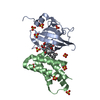
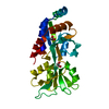
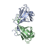
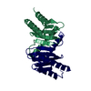
| Deposited unit | 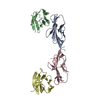
| ||||||||||
|---|---|---|---|---|---|---|---|---|---|---|---|
| 1 | 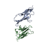
| ||||||||||
| 2 | 
| ||||||||||
| Unit cell |
| ||||||||||
| Details | The biological assembly is each of the individual molecules |
- Components
| #1: Protein | Mass: 12017.655 Da / Num. of mol.: 2 / Fragment: N-TERMINAL ADHESION DOMAIN OF CD2 / Mutation: K61E, F63L, T67A Source method: isolated from a genetically manipulated source Source: (gene. exp.)  Homo sapiens (human) / Plasmid: PET-11A / Production host: Homo sapiens (human) / Plasmid: PET-11A / Production host:  #2: Protein | Mass: 11003.180 Da / Num. of mol.: 2 / Fragment: N-TERMINAL ADHESION DOMAIN OF CD58 / Mutation: F1S, V9K, V21Q, V58K, T85S, L93G Source method: isolated from a genetically manipulated source Source: (gene. exp.)  Homo sapiens (human) / Plasmid: PET-11A / Production host: Homo sapiens (human) / Plasmid: PET-11A / Production host:  |
|---|
-Experimental details
-Experiment
| Experiment | Method:  X-RAY DIFFRACTION / Number of used crystals: 1 X-RAY DIFFRACTION / Number of used crystals: 1 |
|---|
- Sample preparation
| Crystal | Density Matthews: 3.01 Å3/Da / Density % sol: 59.17 % | ||||||||||||||||||||||||||||||||||||||||||
|---|---|---|---|---|---|---|---|---|---|---|---|---|---|---|---|---|---|---|---|---|---|---|---|---|---|---|---|---|---|---|---|---|---|---|---|---|---|---|---|---|---|---|---|
| Crystal grow | Temperature: 298 K / Method: vapor diffusion, hanging drop / pH: 7.5 Details: PEG 400, (NH4)2SO4, NaCl, HEPES, pH 7.5, VAPOR DIFFUSION, HANGING DROP, temperature 298.0K | ||||||||||||||||||||||||||||||||||||||||||
| Crystal | *PLUS Density % sol: 61 % | ||||||||||||||||||||||||||||||||||||||||||
| Crystal grow | *PLUS | ||||||||||||||||||||||||||||||||||||||||||
| Components of the solutions | *PLUS
|
-Data collection
| Diffraction | Mean temperature: 298 K |
|---|---|
| Diffraction source | Source:  ROTATING ANODE / Type: RIGAKU RU300 / Wavelength: 1.5418 ROTATING ANODE / Type: RIGAKU RU300 / Wavelength: 1.5418 |
| Detector | Type: MARRESEARCH / Detector: IMAGE PLATE / Date: Nov 12, 1998 |
| Radiation | Protocol: SINGLE WAVELENGTH / Monochromatic (M) / Laue (L): M / Scattering type: x-ray |
| Radiation wavelength | Wavelength: 1.5418 Å / Relative weight: 1 |
| Reflection | Resolution: 3.2→18 Å / Num. all: 9453 / Num. obs: 8196 / % possible obs: 86.7 % / Observed criterion σ(F): 0 / Observed criterion σ(I): 0 / Redundancy: 5.1 % / Rmerge(I) obs: 0.102 / Net I/σ(I): 9.8 |
| Reflection shell | Resolution: 3.2→3.31 Å / Rmerge(I) obs: 0.278 / Num. unique all: 8196 / % possible all: 87.6 |
| Reflection | *PLUS Num. measured all: 41649 |
| Reflection shell | *PLUS % possible obs: 87.6 % |
- Processing
| Software |
| ||||||||||||||||||||
|---|---|---|---|---|---|---|---|---|---|---|---|---|---|---|---|---|---|---|---|---|---|
| Refinement | Resolution: 3.2→18 Å / σ(F): 2 / Stereochemistry target values: Engh & Huber / Details: Used weighted full matrix least squares procedure.
| ||||||||||||||||||||
| Refinement step | Cycle: LAST / Resolution: 3.2→18 Å
| ||||||||||||||||||||
| Refine LS restraints |
| ||||||||||||||||||||
| Software | *PLUS Name:  X-PLOR / Version: 3.851 / Classification: refinement X-PLOR / Version: 3.851 / Classification: refinement | ||||||||||||||||||||
| Refinement | *PLUS Highest resolution: 3.2 Å / σ(F): 2 / Rfactor obs: 0.223 | ||||||||||||||||||||
| Solvent computation | *PLUS | ||||||||||||||||||||
| Displacement parameters | *PLUS | ||||||||||||||||||||
| Refine LS restraints | *PLUS Type: x_bond_d / Dev ideal: 0.006 |
 Movie
Movie Controller




 PDBj
PDBj

