 Movie
Movie Controller

[English] 日本語
 Yorodumi
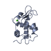
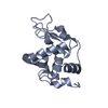
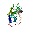
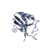
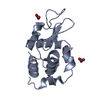
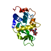
- PDB-1jo0: Structure of HI1333, a Hypothetical Protein from Haemophilus infl... -
+ Open data

- Basic information
| Entry | Database: PDB / ID: 1jo0 | ||||||
|---|---|---|---|---|---|---|---|
| Title | Structure of HI1333, a Hypothetical Protein from Haemophilus influenzae with Structural Similarity to RNA-binding Proteins | ||||||
 Components Components | HYPOTHETICAL PROTEIN HI1333 | ||||||
 Keywords Keywords | STRUCTURAL GENOMICS / unknown function / hypothetical protein / HI1333 / YHBY_HAEIN / Structure 2 Function Project / S2F | ||||||
| Function / homology |  Function and homology information Function and homology informationrRNA 5'-end processing / preribosome binding / ribosomal small subunit assembly / ribosomal large subunit assembly / RNA binding / cytosol Similarity search - Function | ||||||
| Biological species |  Haemophilus influenzae (bacteria) Haemophilus influenzae (bacteria) | ||||||
| Method |  X-RAY DIFFRACTION / SYNCHROTRON / MAD (PT, BR) / Resolution: 1.37 Å X-RAY DIFFRACTION / SYNCHROTRON / MAD (PT, BR) / Resolution: 1.37 Å | ||||||
 Authors Authors | Willis, M.A. / Krajewski, W. / Chalamasetty, V.R. / Reddy, P. / Howard, A. / Herzberg, O. / Structure 2 Function Project (S2F) | ||||||
 Citation Citation | Journal: Proteins / Year: 2002 Title: Structure of HI1333 (YhbY), a putative RNA-binding protein from Haemophilus influenzae Authors: Willis, M.A. / Krajewski, W. / Chalamasetty, V.R. / Reddy, P. / Howard, A. / Herzberg, O. | ||||||
| History |
|
- Structure visualization
| Structure viewer | Molecule: MolmilJmol/JSmol |
|---|
- Downloads & links
-Download
| PDBx/mmCIF format | 1jo0.cif.gz | 103.9 KB | Display | PDBx/mmCIF format |
|---|---|---|---|---|
| PDB format | pdb1jo0.ent.gz | 80.7 KB | Display | PDB format |
| PDBx/mmJSON format | 1jo0.json.gz | Tree view | PDBx/mmJSON format | |
| Others |  Other downloads Other downloads |
-Validation report
| Arichive directory | https://data.pdbj.org/pub/pdb/validation_reports/jo/1jo0ftp://data.pdbj.org/pub/pdb/validation_reports/jo/1jo0 | HTTPS FTP |
|---|
-Related structure data
| Similar structure data | |
|---|---|
| Other databases |


-Links
 PDBj
PDBj- Assembly
| Deposited unit | 
| ||||||||
|---|---|---|---|---|---|---|---|---|---|
| 1 | 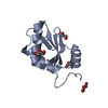
| ||||||||
| 2 | 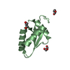
| ||||||||
| Unit cell |
| ||||||||
| Details | Dynamic Light Scattering (DLS) indicates the protein is a monomer in solution. There are two monomers in the asymmetric unit. |
-Components
| #1: Protein | Mass: 10810.617 Da / Num. of mol.: 2 Source method: isolated from a genetically manipulated source Source: (gene. exp.) Haemophilus influenzae (bacteria) / Gene: HI1333 / Plasmid: pRE1 / Production host: #2: Chemical | ChemComp-GOL /   Mass: 92.094 Da / Num. of mol.: 7 / Source method: obtained synthetically / Formula: C3H8O3 Mass: 92.094 Da / Num. of mol.: 7 / Source method: obtained synthetically / Formula: C3H8O3#3: Water | ChemComp-HOH / |  Mass: 18.015 Da / Num. of mol.: 264 / Source method: isolated from a natural source / Formula: H2O Mass: 18.015 Da / Num. of mol.: 264 / Source method: isolated from a natural source / Formula: H2O |
|---|
-Experimental details
-Experiment
| Experiment | Method: X-RAY DIFFRACTION / Number of used crystals: 1 |
|---|
- Sample preparation
| Crystal | Density Matthews: 1.77 Å3/Da / Density % sol: 41.99 % | ||||||||||||||||||||||||||||
|---|---|---|---|---|---|---|---|---|---|---|---|---|---|---|---|---|---|---|---|---|---|---|---|---|---|---|---|---|---|
| Crystal grow | Temperature: 298 K / Method: vapor diffusion, hanging drop / pH: 8 Details: 28-30% PEG4000, 100mM Tris pH 8.0, 10mM CaCl2, + 1.5% heptane-1,2,3-triol added to drop, VAPOR DIFFUSION, HANGING DROP, temperature 298K | ||||||||||||||||||||||||||||
| Crystal grow | *PLUS pH: 7 / Method: microdialysis | ||||||||||||||||||||||||||||
| Components of the solutions | *PLUS
|
-Data collection
| Diffraction | Mean temperature: 100 K |
|---|---|
| Diffraction source | Source: SYNCHROTRON / Site: APS  / Beamline: 17-BM / Wavelength: 0.9184 / Wavelength: 0.9184 Å / Beamline: 17-BM / Wavelength: 0.9184 / Wavelength: 0.9184 Å |
| Detector | Type: MARRESEARCH / Detector: CCD / Date: Mar 10, 2001 / Details: PT/PD-COATED ULE MIRROR |
| Radiation | Monochromator: SI(111) / Protocol: SINGLE WAVELENGTH / Monochromatic (M) / Laue (L): M / Scattering type: x-ray |
| Radiation wavelength | Wavelength: 0.9184 Å / Relative weight: 1 |
| Reflection | Resolution: 1.37→25.9 Å / Num. all: 38052 / Num. obs: 38052 / % possible obs: 99.9 % / Observed criterion σ(I): -3 / Redundancy: 6.47 % / Rmerge(I) obs: 0.0308 / Net I/σ(I): 26.79 |
| Reflection shell | Resolution: 1.37→1.4 Å / Redundancy: 3.63 % / Rmerge(I) obs: 0.2705 / Mean I/σ(I) obs: 3.06 / Num. unique all: 2391 / % possible all: 100 |
| Reflection | *PLUS Num. obs: 38069 / Num. measured all: 246633 / Rmerge(I) obs: 0.031 |
| Reflection shell | *PLUS Lowest resolution: 1.42 Å / % possible obs: 100 % / Rmerge(I) obs: 0.271 |
- Processing
| Software |
| ||||||||||||||||||||||||||||||||||||
|---|---|---|---|---|---|---|---|---|---|---|---|---|---|---|---|---|---|---|---|---|---|---|---|---|---|---|---|---|---|---|---|---|---|---|---|---|---|
| Refinement | Method to determine structure: MAD (PT, BR) / Resolution: 1.37→25.9 Å / Num. parameters: 16757 / Num. restraintsaints: 20338 / Cross valid method: FREE R / σ(I): -3 / Stereochemistry target values: ENGH AND HUBER
| ||||||||||||||||||||||||||||||||||||
| Refine analyze | Num. disordered residues: 14 / Occupancy sum hydrogen: 1525 / Occupancy sum non hydrogen: 1771.5 | ||||||||||||||||||||||||||||||||||||
| Refinement step | Cycle: LAST / Resolution: 1.37→25.9 Å
| ||||||||||||||||||||||||||||||||||||
| Refine LS restraints |
| ||||||||||||||||||||||||||||||||||||
| Software | *PLUS Name: SHELXL / Version: 97 / Classification: refinement | ||||||||||||||||||||||||||||||||||||
| Refinement | *PLUS % reflection Rfree: 8 % / Rfactor Rfree: 0.221 / Rfactor Rwork: 0.138 | ||||||||||||||||||||||||||||||||||||
| Solvent computation | *PLUS | ||||||||||||||||||||||||||||||||||||
| Displacement parameters | *PLUS | ||||||||||||||||||||||||||||||||||||
| Refine LS restraints | *PLUS
| ||||||||||||||||||||||||||||||||||||
| LS refinement shell | *PLUS Rfactor Rfree: 0.213 / Rfactor Rwork: 0.132 |
