 Movie
Movie Controller

[English] 日本語
 Yorodumi
- PDB-7cyv: Crystal structure of FD20, a neutralizing single-chain variable f... -
+ Open data

- Basic information
| Entry | Database: PDB / ID: 7cyv | ||||||
|---|---|---|---|---|---|---|---|
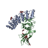
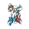
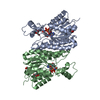
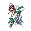
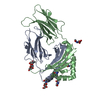
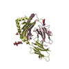
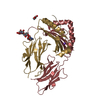
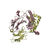
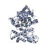
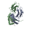
| Title | Crystal structure of FD20, a neutralizing single-chain variable fragment (scFv) in complex with SARS-CoV-2 Spike receptor-binding domain (RBD) | ||||||
 Components Components |
| ||||||
 Keywords Keywords | VIRAL PROTEIN / coronavirus / Covid-19 / nCoV-2019 / neutralizing antibody / receptor-binding domain / SARS-CoV-2 / scFv / single-chain variable fragment / Spike | ||||||
| Function / homology |  Function and homology information Function and homology informationsymbiont-mediated disruption of host tissue / Maturation of spike protein / Translation of Structural Proteins / Virion Assembly and Release / host cell surface / host extracellular region / symbiont-mediated-mediated suppression of host tetherin activity / Induction of Cell-Cell Fusion / structural constituent of virion / positive regulation of viral entry into host cell ...symbiont-mediated disruption of host tissue / Maturation of spike protein / Translation of Structural Proteins / Virion Assembly and Release / host cell surface / host extracellular region / symbiont-mediated-mediated suppression of host tetherin activity / Induction of Cell-Cell Fusion / structural constituent of virion / positive regulation of viral entry into host cell / membrane fusion / host cell endoplasmic reticulum-Golgi intermediate compartment membrane / Attachment and Entry / entry receptor-mediated virion attachment to host cell / receptor-mediated virion attachment to host cell / host cell surface receptor binding / symbiont-mediated suppression of host innate immune response / endocytosis involved in viral entry into host cell / receptor ligand activity / fusion of virus membrane with host plasma membrane / fusion of virus membrane with host endosome membrane / viral envelope / symbiont entry into host cell / virion attachment to host cell / host cell plasma membrane / SARS-CoV-2 activates/modulates innate and adaptive immune responses / virion membrane / membrane / identical protein binding / plasma membrane Similarity search - Function | ||||||
| Biological species |  Homo sapiens (human) Homo sapiens (human)  Severe acute respiratory syndrome coronavirus 2 Severe acute respiratory syndrome coronavirus 2 | ||||||
| Method |  X-RAY DIFFRACTION / SYNCHROTRON / MOLECULAR REPLACEMENT / Resolution: 3.13 Å X-RAY DIFFRACTION / SYNCHROTRON / MOLECULAR REPLACEMENT / Resolution: 3.13 Å | ||||||
 Authors Authors | Li, Y. / Li, T. / Lai, Y. / Cai, H. / Yao, H. / Li, D. | ||||||
| Funding support |  China, 1items China, 1items
| ||||||
 Citation Citation | Journal: Embo Mol Med / Year: 2021 Title: Uncovering a conserved vulnerability site in SARS-CoV-2 by a human antibody. Authors: Li, T. / Cai, H. / Zhao, Y. / Li, Y. / Lai, Y. / Yao, H. / Liu, L.D. / Sun, Z. / van Vlissingen, M.F. / Kuiken, T. / GeurtsvanKessel, C.H. / Zhang, N. / Zhou, B. / Lu, L. / Gong, Y. / Qin, W. ...Authors: Li, T. / Cai, H. / Zhao, Y. / Li, Y. / Lai, Y. / Yao, H. / Liu, L.D. / Sun, Z. / van Vlissingen, M.F. / Kuiken, T. / GeurtsvanKessel, C.H. / Zhang, N. / Zhou, B. / Lu, L. / Gong, Y. / Qin, W. / Mondal, M. / Duan, B. / Xu, S. / Richard, A.S. / Raoul, H. / Chen, J. / Xu, C. / Wu, L. / Zhou, H. / Huang, Z. / Zhang, X. / Li, J. / Wang, Y. / Bi, Y. / Rockx, B. / Chen, J. / Meng, F.L. / Lavillette, D. / Li, D. | ||||||
| History |
|
- Structure visualization
| Structure viewer | Molecule: MolmilJmol/JSmol |
|---|
- Downloads & links
-Download
| PDBx/mmCIF format | 7cyv.cif.gz | 107.1 KB | Display | PDBx/mmCIF format |
|---|---|---|---|---|
| PDB format | pdb7cyv.ent.gz | 65.3 KB | Display | PDB format |
| PDBx/mmJSON format | 7cyv.json.gz | Tree view | PDBx/mmJSON format | |
| Others |  Other downloads Other downloads |
-Validation report
| Arichive directory | https://data.pdbj.org/pub/pdb/validation_reports/cy/7cyvftp://data.pdbj.org/pub/pdb/validation_reports/cy/7cyv | HTTPS FTP |
|---|
-Related structure data


-Links
 PDBj
PDBj


- Assembly
| Deposited unit | 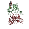
| ||||||||||||
|---|---|---|---|---|---|---|---|---|---|---|---|---|---|
| 1 |
| ||||||||||||
| Unit cell |
|
-Components
| #1: Antibody | Mass: 29613.533 Da / Num. of mol.: 1 Source method: isolated from a genetically manipulated source Details: The fusion protein of tag (GSSS), heavy chain variable region of the scFv FD20 (residues 0-125), linker (GGGSGGGGSGGGGSS), light chain variable region of the scFv FD20 (residues 1002-1129), and tag (HHHHHH) Source: (gene. exp.) Homo sapiens (human) / Production host:  |
|---|---|
| #2: Protein | Mass: 23827.623 Da / Num. of mol.: 1 / Fragment: receptor-binding domain (RBD) Source method: isolated from a genetically manipulated source Source: (gene. exp.) Severe acute respiratory syndrome coronavirus 2Gene: S, 2 / Cell line (production host): High Five / Production host:  Trichoplusia ni (cabbage looper) / References: UniProt: P0DTC2 Trichoplusia ni (cabbage looper) / References: UniProt: P0DTC2 |
| #3: Polysaccharide | beta-D-mannopyranose-(1-4)-2-acetamido-2-deoxy-beta-D-glucopyranose-(1-4)-[alpha-L-fucopyranose-(1- ...beta-D-mannopyranose-(1-4)-2-acetamido-2-deoxy-beta-D-glucopyranose-(1-4)-[alpha-L-fucopyranose-(1-3)][alpha-L-fucopyranose-(1-6)]2-acetamido-2-deoxy-beta-D-glucopyranose Type: oligosaccharide / Mass: 878.823 Da / Num. of mol.: 1 Source method: isolated from a genetically manipulated source |
| Has ligand of interest | Y |
| Has protein modification | Y |
-Experimental details
-Experiment
| Experiment | Method: X-RAY DIFFRACTION / Number of used crystals: 1 |
|---|
- Sample preparation
| Crystal | Density Matthews: 2.6 Å3/Da / Density % sol: 52.76 % |
|---|---|
| Crystal grow | Temperature: 293 K / Method: vapor diffusion, sitting drop / pH: 5.5 Details: 0.2 M lithium sulfate monohydrate, 0.1 M Bis-Tris pH 5.5, 25% w/v polyethylene glycol 3350 |
-Data collection
| Diffraction | Mean temperature: 100 K / Serial crystal experiment: N |
|---|---|
| Diffraction source | Source: SYNCHROTRON / Site: SSRF / Beamline: BL18U1 / Wavelength: 0.97915 Å |
| Detector | Type: PSI PILATUS 6M / Detector: PIXEL / Date: Jul 3, 2020 |
| Radiation | Protocol: SINGLE WAVELENGTH / Monochromatic (M) / Laue (L): M / Scattering type: x-ray |
| Radiation wavelength | Wavelength: 0.97915 Å / Relative weight: 1 |
| Reflection | Resolution: 3.13→46.43 Å / Num. obs: 9847 / % possible obs: 98.89 % / Redundancy: 5.2 % / Biso Wilson estimate: 65.25 Å2 / CC1/2: 0.979 / Rmerge(I) obs: 0.2724 / Rpim(I) all: 0.1322 / Net I/σ(I): 5.65 |
| Reflection shell | Resolution: 3.13→3.24 Å / Redundancy: 5.3 % / Rmerge(I) obs: 1.125 / Mean I/σ(I) obs: 1.5 / Num. unique obs: 927 / CC1/2: 0.791 / Rpim(I) all: 0.5387 / % possible all: 94.68 |
- Processing
| Software |
| ||||||||||||||||||||||||||||
|---|---|---|---|---|---|---|---|---|---|---|---|---|---|---|---|---|---|---|---|---|---|---|---|---|---|---|---|---|---|
| Refinement | Method to determine structure: MOLECULAR REPLACEMENT Starting model: 6MOJ, 5C6W Resolution: 3.13→46.43 Å / SU ML: 0.5228 / Cross valid method: FREE R-VALUE / σ(F): 1.35 / Phase error: 31.1232 Stereochemistry target values: GeoStd + Monomer Library + CDL v1.2
| ||||||||||||||||||||||||||||
| Solvent computation | Shrinkage radii: 0.9 Å / VDW probe radii: 1.11 Å / Solvent model: FLAT BULK SOLVENT MODEL | ||||||||||||||||||||||||||||
| Displacement parameters | Biso mean: 61.91 Å2 | ||||||||||||||||||||||||||||
| Refinement step | Cycle: LAST / Resolution: 3.13→46.43 Å
| ||||||||||||||||||||||||||||
| Refine LS restraints |
| ||||||||||||||||||||||||||||
| LS refinement shell |
|
