Movie
Movie Controller


+ Open data

- Basic information
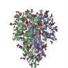
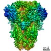
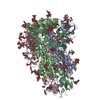
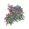
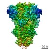
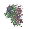
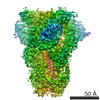
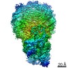
| Entry | Database: PDB / ID: 7cyc | |||||||||
|---|---|---|---|---|---|---|---|---|---|---|
| Title | Cryo-EM structures of Alphacoronavirus spike glycoprotein | |||||||||
 Components Components | Spike glycoprotein | |||||||||
 Keywords Keywords | STRUCTURAL PROTEIN / Alphacoronavirus / spike glycoprotein | |||||||||
| Function / homology |  Function and homology information Function and homology informationhost cell endoplasmic reticulum-Golgi intermediate compartment membrane / receptor-mediated virion attachment to host cell / endocytosis involved in viral entry into host cell / fusion of virus membrane with host plasma membrane / fusion of virus membrane with host endosome membrane / viral envelope / virion membrane / membrane Similarity search - Function | |||||||||
| Biological species |  Human coronavirus 229E Human coronavirus 229E | |||||||||
| Method | ELECTRON MICROSCOPY / single particle reconstruction / cryo EM / Resolution: 3.21 Å | |||||||||
 Authors Authors | Song, X. / Shi, Y. / Ding, W. / Liu, Z.J. / Peng, G. | |||||||||
| Funding support |  China, 2items China, 2items
| |||||||||
 Citation Citation | Journal: Nat Commun / Year: 2021 Title: Cryo-EM analysis of the HCoV-229E spike glycoprotein reveals dynamic prefusion conformational changes. Authors: Xiyong Song / Yuejun Shi / Wei Ding / Tongxin Niu / Limeng Sun / Yubei Tan / Yong Chen / Jiale Shi / Qiqi Xiong / Xiaojun Huang / Shaobo Xiao / Yanping Zhu / Chongyun Cheng / Zhen F Fu / Zhi- ...Authors: Xiyong Song / Yuejun Shi / Wei Ding / Tongxin Niu / Limeng Sun / Yubei Tan / Yong Chen / Jiale Shi / Qiqi Xiong / Xiaojun Huang / Shaobo Xiao / Yanping Zhu / Chongyun Cheng / Zhen F Fu / Zhi-Jie Liu / Guiqing Peng /  Abstract: Coronaviruses spike (S) glycoproteins mediate viral entry into host cells by binding to host receptors. However, how the S1 subunit undergoes conformational changes for receptor recognition has not ...Coronaviruses spike (S) glycoproteins mediate viral entry into host cells by binding to host receptors. However, how the S1 subunit undergoes conformational changes for receptor recognition has not been elucidated in Alphacoronavirus. Here, we report the cryo-EM structures of the HCoV-229E S trimer in prefusion state with two conformations. The activated conformation may pose the potential exposure of the S1-RBDs by decreasing of the interaction area between the S1-RBDs and the surrounding S1-NTDs and S1-RBDs compared to the closed conformation. Furthermore, structural comparison of our structures with the previously reported HCoV-229E S structure showed that the S trimers trended to open the S2 subunit from the closed conformation to open conformation, which could promote the transition from pre- to postfusion. Our results provide insights into the mechanisms involved in S glycoprotein-mediated Alphacoronavirus entry and have implications for vaccine and therapeutic antibody design. | |||||||||
| History |
|
- Structure visualization
| Movie |
Movie viewer |
|---|---|
| Structure viewer | Molecule: MolmilJmol/JSmol |
- Downloads & links
-Download
| PDBx/mmCIF format | 7cyc.cif.gz | 517.6 KB | Display | PDBx/mmCIF format |
|---|---|---|---|---|
| PDB format | pdb7cyc.ent.gz | 420.8 KB | Display | PDB format |
| PDBx/mmJSON format | 7cyc.json.gz | Tree view | PDBx/mmJSON format | |
| Others |  Other downloads Other downloads |
-Validation report
| Arichive directory | https://data.pdbj.org/pub/pdb/validation_reports/cy/7cycftp://data.pdbj.org/pub/pdb/validation_reports/cy/7cyc | HTTPS FTP |
|---|
-Related structure data
| Related structure data | 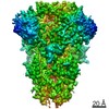 30496MC 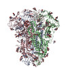 7cydC M: map data used to model this data C: citing same article ( |
|---|---|
| Similar structure data |

-Links
 PDBj
PDBj- Assembly
| Deposited unit | 
|
|---|---|
| 1 |
|
-Components
| #1: Protein | Mass: 122402.008 Da / Num. of mol.: 3 Source method: isolated from a genetically manipulated source Source: (gene. exp.) Human coronavirus 229E / Gene: S, 2Production host: Insect cell expression vector pTIE1 (others) References: UniProt: P15423 #2: Polysaccharide | Source method: isolated from a genetically manipulated source #3: Polysaccharide | beta-D-mannopyranose-(1-4)-2-acetamido-2-deoxy-beta-D-glucopyranose-(1-4)-2-acetamido-2-deoxy-beta- ...beta-D-mannopyranose-(1-4)-2-acetamido-2-deoxy-beta-D-glucopyranose-(1-4)-2-acetamido-2-deoxy-beta-D-glucopyranose Source method: isolated from a genetically manipulated source #4: Polysaccharide | Source method: isolated from a genetically manipulated source #5: Sugar | ChemComp-NAG /   Type: D-saccharide, beta linking / Mass: 221.208 Da / Num. of mol.: 48 / Source method: obtained synthetically / Formula: C8H15NO6 / Feature type: SUBJECT OF INVESTIGATION Type: D-saccharide, beta linking / Mass: 221.208 Da / Num. of mol.: 48 / Source method: obtained synthetically / Formula: C8H15NO6 / Feature type: SUBJECT OF INVESTIGATIONHas ligand of interest | Y | Has protein modification | Y | |
|---|
-Experimental details
-Experiment
| Experiment | Method: ELECTRON MICROSCOPY |
|---|---|
| EM experiment | Aggregation state: PARTICLE / 3D reconstruction method: single particle reconstruction |
- Sample preparation
| Component | Name: HCoV-229E spike trimer / Type: COMPLEX / Entity ID: #1 / Source: RECOMBINANT | |||||||||||||||
|---|---|---|---|---|---|---|---|---|---|---|---|---|---|---|---|---|
| Source (natural) | Organism: Human coronavirus 229E | |||||||||||||||
| Source (recombinant) | Organism: eukar | |||||||||||||||
| Buffer solution | pH: 7.2 | |||||||||||||||
| Buffer component |
| |||||||||||||||
| Specimen | Conc.: 0.75 mg/ml / Embedding applied: NO / Shadowing applied: NO / Staining applied: NO / Vitrification applied: YES | |||||||||||||||
| Specimen support | Grid type: Quantifoil R1.2/1.3 | |||||||||||||||
| Vitrification | Instrument: FEI VITROBOT MARK IV / Cryogen name: ETHANE / Humidity: 100 % / Chamber temperature: 277 K |
- Electron microscopy imaging
| Experimental equipment |  Model: Titan Krios / Image courtesy: FEI Company |
|---|---|
| Microscopy | Model: FEI TITAN KRIOS |
| Electron gun | Electron source:  FIELD EMISSION GUN / Accelerating voltage: 300 kV / Illumination mode: FLOOD BEAM FIELD EMISSION GUN / Accelerating voltage: 300 kV / Illumination mode: FLOOD BEAM |
| Electron lens | Mode: BRIGHT FIELD / Nominal magnification: 18000 X / Nominal defocus max: 3000 nm / Nominal defocus min: 2000 nm / Calibrated defocus min: 2000 nm / Calibrated defocus max: 3000 nm / Cs: 2.7 mm / C2 aperture diameter: 100 µm / Alignment procedure: COMA FREE |
| Specimen holder | Cryogen: NITROGEN / Specimen holder model: FEI TITAN KRIOS AUTOGRID HOLDER / Temperature (max): 80 K / Temperature (min): 80 K / Residual tilt: 5 mradians |
| Image recording | Electron dose: 60 e/Å2 / Detector mode: SUPER-RESOLUTION / Film or detector model: GATAN K2 SUMMIT (4k x 4k) |
| Image scans | Scanner model: OTHER |
- Processing
| Software | Name: PHENIX / Version: 1.18_3855: / Classification: refinement | |||||||||||||||||||||||||||||||||||||||||||||
|---|---|---|---|---|---|---|---|---|---|---|---|---|---|---|---|---|---|---|---|---|---|---|---|---|---|---|---|---|---|---|---|---|---|---|---|---|---|---|---|---|---|---|---|---|---|---|
| EM software |
| |||||||||||||||||||||||||||||||||||||||||||||
| CTF correction | Type: PHASE FLIPPING AND AMPLITUDE CORRECTION | |||||||||||||||||||||||||||||||||||||||||||||
| Particle selection | Num. of particles selected: 403347 | |||||||||||||||||||||||||||||||||||||||||||||
| 3D reconstruction | Resolution: 3.21 Å / Resolution method: FSC 0.143 CUT-OFF / Num. of particles: 36846 / Symmetry type: POINT | |||||||||||||||||||||||||||||||||||||||||||||
| Atomic model building | B value: 100 / Protocol: FLEXIBLE FIT / Space: REAL / Target criteria: Correlation coefficient | |||||||||||||||||||||||||||||||||||||||||||||
| Atomic model building | PDB-ID: 5SZS Pdb chain-ID: A / Accession code: 5SZS / Pdb chain residue range: 17-1012 / Source name: PDB / Type: experimental model | |||||||||||||||||||||||||||||||||||||||||||||
| Refine LS restraints |
|