 Movie
Movie Controller

[English] 日本語
 Yorodumi
- PDB-7cx0: Crystal structure of a tyrosine decarboxylase from Enterococcus f... -
+ Open data

- Basic information
| Entry | Database: PDB / ID: 7cx0 | ||||||
|---|---|---|---|---|---|---|---|
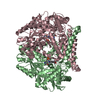
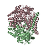
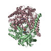
| Title | Crystal structure of a tyrosine decarboxylase from Enterococcus faecalis in complex with the cofactor PLP and inhibitor carbidopa | ||||||
 Components Components | Decarboxylase | ||||||
 Keywords Keywords | LYASE / Catalytic / binding pocket / tyrosine decarboxylase | ||||||
| Function / homology |  Function and homology information Function and homology informationdiaminobutyrate decarboxylase / diaminobutyrate decarboxylase activity / tyrosine decarboxylase / tyrosine decarboxylase activity / aromatic-L-amino-acid decarboxylase activity / carboxylic acid metabolic process / pyridoxal phosphate binding Similarity search - Function | ||||||
| Biological species |   Enterococcus faecalis (bacteria) Enterococcus faecalis (bacteria) | ||||||
| Method |  X-RAY DIFFRACTION / SYNCHROTRON / MOLECULAR REPLACEMENT / Resolution: 2.66 Å X-RAY DIFFRACTION / SYNCHROTRON / MOLECULAR REPLACEMENT / Resolution: 2.66 Å | ||||||
 Authors Authors | Yu, X. / Gong, M. / Huang, J. / Liu, W. / Chen, C. / Guo, R. | ||||||
 Citation Citation | Journal: to be published Title: Crystal structure of a tyrosine decarboxylase from Enterococcus faecalis in complex with the cofactor PLP and inhibitor carbidopa Authors: Yu, X. / Gong, M. / Huang, J. / Liu, W. / Chen, C. / Guo, R. | ||||||
| History |
|
- Structure visualization
| Structure viewer | Molecule: MolmilJmol/JSmol |
|---|
- Downloads & links
-Download
| PDBx/mmCIF format | 7cx0.cif.gz | 357.6 KB | Display | PDBx/mmCIF format |
|---|---|---|---|---|
| PDB format | pdb7cx0.ent.gz | 289.7 KB | Display | PDB format |
| PDBx/mmJSON format | 7cx0.json.gz | Tree view | PDBx/mmJSON format | |
| Others |  Other downloads Other downloads |
-Validation report
| Arichive directory | https://data.pdbj.org/pub/pdb/validation_reports/cx/7cx0ftp://data.pdbj.org/pub/pdb/validation_reports/cx/7cx0 | HTTPS FTP |
|---|
-Related structure data
| Related structure data |  5hsiS S: Starting model for refinement |
|---|---|
| Similar structure data |


-Links
 PDBj
PDBj- Assembly
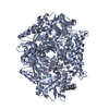
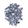
| Deposited unit | 
| ||||||||||||||||||||||||||||||||||||||||||||||||||||||||||||||||||||||||||||
|---|---|---|---|---|---|---|---|---|---|---|---|---|---|---|---|---|---|---|---|---|---|---|---|---|---|---|---|---|---|---|---|---|---|---|---|---|---|---|---|---|---|---|---|---|---|---|---|---|---|---|---|---|---|---|---|---|---|---|---|---|---|---|---|---|---|---|---|---|---|---|---|---|---|---|---|---|---|
| 1 | 
| ||||||||||||||||||||||||||||||||||||||||||||||||||||||||||||||||||||||||||||
| 2 | 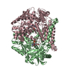
| ||||||||||||||||||||||||||||||||||||||||||||||||||||||||||||||||||||||||||||
| Unit cell |
| ||||||||||||||||||||||||||||||||||||||||||||||||||||||||||||||||||||||||||||
| Noncrystallographic symmetry (NCS) | NCS domain:
NCS domain segments:
|