 Movie
Movie Controller


+ Open data

- Basic information
| Entry | Database: PDB / ID: 7csv | ||||||
|---|---|---|---|---|---|---|---|
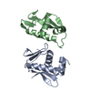
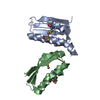
| Title | Pseudomonas aeruginosa antitoxin HigA | ||||||
 Components Components | HTH cro/C1-type domain-containing protein | ||||||
 Keywords Keywords | ANTITOXIN / dimer | ||||||
| Function / homology | Toxin-antitoxin system, antidote protein, HigA / Helix-turn-helix / Helix-turn-helix XRE-family like proteins / Cro/C1-type HTH domain profile. / Cro/C1-type helix-turn-helix domain / Lambda repressor-like, DNA-binding domain superfamily / regulation of DNA-templated transcription / DNA binding / HTH cro/C1-type domain-containing protein Function and homology information Function and homology information | ||||||
| Biological species |  Pseudomonas aeruginosa PAO1 (bacteria) Pseudomonas aeruginosa PAO1 (bacteria) | ||||||
| Method |  X-RAY DIFFRACTION / SYNCHROTRON / MOLECULAR REPLACEMENT / Resolution: 1.71 Å X-RAY DIFFRACTION / SYNCHROTRON / MOLECULAR REPLACEMENT / Resolution: 1.71 Å | ||||||
 Authors Authors | Song, Y.J. / Luo, G.H. / Bao, R. | ||||||
 Citation Citation | Journal: Environ.Microbiol. / Year: 2021 Title: Pseudomonas aeruginosa antitoxin HigA functions as a diverse regulatory factor by recognizing specific pseudopalindromic DNA motifs. Authors: Song, Y. / Luo, G. / Zhu, Y. / Li, T. / Li, C. / He, L. / Zhao, N. / Zhao, C. / Yang, J. / Huang, Q. / Mu, X. / Tang, X. / Kang, M. / Wu, S. / He, Y. / Bao, R. | ||||||
| History |
|
- Structure visualization
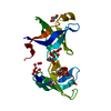
| Structure viewer | Molecule: MolmilJmol/JSmol |
|---|
- Downloads & links
-Download
| PDBx/mmCIF format | 7csv.cif.gz | 64.3 KB | Display | PDBx/mmCIF format |
|---|---|---|---|---|
| PDB format | pdb7csv.ent.gz | 38.4 KB | Display | PDB format |
| PDBx/mmJSON format | 7csv.json.gz | Tree view | PDBx/mmJSON format | |
| Others |  Other downloads Other downloads |
-Validation report
| Arichive directory | https://data.pdbj.org/pub/pdb/validation_reports/cs/7csvftp://data.pdbj.org/pub/pdb/validation_reports/cs/7csv | HTTPS FTP |
|---|
-Related structure data
| Related structure data |  7cswC  7csyC  6f8hS S: Starting model for refinement C: citing same article ( |
|---|---|
| Similar structure data |


-Links
 PDBj
PDBj


- Assembly
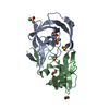
| Deposited unit | 
| ||||||||||||
|---|---|---|---|---|---|---|---|---|---|---|---|---|---|
| 1 |
| ||||||||||||
| Unit cell |
| ||||||||||||
| Components on special symmetry positions |
|
-Components
| #1: Protein | Mass: 11259.780 Da / Num. of mol.: 2 Source method: isolated from a genetically manipulated source Source: (gene. exp.) Pseudomonas aeruginosa PAO1 (bacteria) / Strain: PAO1 / Gene: PA4674 / Production host: #2: Water | ChemComp-HOH / |  Mass: 18.015 Da / Num. of mol.: 135 / Source method: isolated from a natural source / Formula: H2O Mass: 18.015 Da / Num. of mol.: 135 / Source method: isolated from a natural source / Formula: H2O |
|---|
-Experimental details
-Experiment
| Experiment | Method: X-RAY DIFFRACTION / Number of used crystals: 1 |
|---|
- Sample preparation
| Crystal | Density Matthews: 1.94 Å3/Da / Density % sol: 36.64 % |
|---|---|
| Crystal grow | Temperature: 290 K / Method: vapor diffusion, sitting drop / Details: 0.2M MgCl2, 0.1M Tris pH 8.5, 20% PEG8000 |
-Data collection
| Diffraction | Mean temperature: 100 K / Serial crystal experiment: N |
|---|---|
| Diffraction source | Source: SYNCHROTRON / Site: SSRF  / Beamline: BL18U1 / Wavelength: 0.9793 Å / Beamline: BL18U1 / Wavelength: 0.9793 Å |
| Detector | Type: ADSC QUANTUM 315r / Detector: CCD / Date: Aug 1, 2019 |
| Radiation | Protocol: SINGLE WAVELENGTH / Monochromatic (M) / Laue (L): M / Scattering type: x-ray |
| Radiation wavelength | Wavelength: 0.9793 Å / Relative weight: 1 |
| Reflection | Resolution: 1.707→19.54 Å / Num. obs: 33999 / % possible obs: 99.9 % / Redundancy: 33.8 % / Biso Wilson estimate: 15.87 Å2 / CC1/2: 0.996 / Rmerge(I) obs: 0.116 / Net I/σ(I): 6.4 |
| Reflection shell | Resolution: 1.707→1.768 Å / Num. unique obs: 1875 / CC1/2: 0.993 |
- Processing
| Software |
| |||||||||||||||||||||||||||||||||||||||||||||||||||||||||||||||||||||||||||||||||||||||||||||||||||||||||||||||||||||||||||||||||||||||||||||||||||||||||||||||||||||||||||||||
|---|---|---|---|---|---|---|---|---|---|---|---|---|---|---|---|---|---|---|---|---|---|---|---|---|---|---|---|---|---|---|---|---|---|---|---|---|---|---|---|---|---|---|---|---|---|---|---|---|---|---|---|---|---|---|---|---|---|---|---|---|---|---|---|---|---|---|---|---|---|---|---|---|---|---|---|---|---|---|---|---|---|---|---|---|---|---|---|---|---|---|---|---|---|---|---|---|---|---|---|---|---|---|---|---|---|---|---|---|---|---|---|---|---|---|---|---|---|---|---|---|---|---|---|---|---|---|---|---|---|---|---|---|---|---|---|---|---|---|---|---|---|---|---|---|---|---|---|---|---|---|---|---|---|---|---|---|---|---|---|---|---|---|---|---|---|---|---|---|---|---|---|---|---|---|---|---|
| Refinement | Method to determine structure: MOLECULAR REPLACEMENT Starting model: 6F8H Resolution: 1.71→19.54 Å / SU ML: 0.195 / Cross valid method: FREE R-VALUE / σ(F): 1.68 / Phase error: 22.5474 / Stereochemistry target values: GeoStd + Monomer Library Details: SF FILE CONTAINS FRIEDEL PAIRS UNDER I_MINUS AND I_PLUS COLUMNS.
| |||||||||||||||||||||||||||||||||||||||||||||||||||||||||||||||||||||||||||||||||||||||||||||||||||||||||||||||||||||||||||||||||||||||||||||||||||||||||||||||||||||||||||||||
| Solvent computation | Shrinkage radii: 0.9 Å / VDW probe radii: 1.11 Å / Solvent model: FLAT BULK SOLVENT MODEL | |||||||||||||||||||||||||||||||||||||||||||||||||||||||||||||||||||||||||||||||||||||||||||||||||||||||||||||||||||||||||||||||||||||||||||||||||||||||||||||||||||||||||||||||
| Displacement parameters | Biso mean: 18.98 Å2 | |||||||||||||||||||||||||||||||||||||||||||||||||||||||||||||||||||||||||||||||||||||||||||||||||||||||||||||||||||||||||||||||||||||||||||||||||||||||||||||||||||||||||||||||
| Refinement step | Cycle: LAST / Resolution: 1.71→19.54 Å
| |||||||||||||||||||||||||||||||||||||||||||||||||||||||||||||||||||||||||||||||||||||||||||||||||||||||||||||||||||||||||||||||||||||||||||||||||||||||||||||||||||||||||||||||
| Refine LS restraints |
| |||||||||||||||||||||||||||||||||||||||||||||||||||||||||||||||||||||||||||||||||||||||||||||||||||||||||||||||||||||||||||||||||||||||||||||||||||||||||||||||||||||||||||||||
| LS refinement shell |
|
