 Movie
Movie Controller

[English] 日本語
 Yorodumi
- PDB-7cnt: Structure of 2,5-dihydroxypridine Dioxygenase from Pseudomonas pu... -
+ Open data

- Basic information
| Entry | Database: PDB / ID: 7cnt | ||||||
|---|---|---|---|---|---|---|---|
| Title | Structure of 2,5-dihydroxypridine Dioxygenase from Pseudomonas putida KT2440 in complex with product N-formylmaleamic acid formed via in crystallo reaction with 2,5-dihydroxypridine | ||||||
 Components Components | 2,5-dihydroxypyridine 5,6-dioxygenase | ||||||
 Keywords Keywords | OXIDOREDUCTASE / dihydroxypridine / formylmaleamic acid | ||||||
| Function / homology | 2,5-dihydroxypyridine 5,6-dioxygenase / 2,5-dihydroxypyridine 5,6-dioxygenase activity / : / nicotinate catabolic process / metal ion binding / : / 2,5-dihydroxypyridine 5,6-dioxygenase Function and homology information Function and homology information | ||||||
| Biological species |  Pseudomonas putida KT2440 (bacteria) Pseudomonas putida KT2440 (bacteria) | ||||||
| Method |  X-RAY DIFFRACTION / SYNCHROTRON / SAD / Resolution: 2.28 Å X-RAY DIFFRACTION / SYNCHROTRON / SAD / Resolution: 2.28 Å | ||||||
 Authors Authors | Liu, G.Q. / Tang, H.Z. | ||||||
| Funding support |  China, 1items China, 1items
| ||||||
 Citation Citation | Journal: Nat Commun / Year: 2020 Title: 2,5-dihydroxypridine Dioxygenase in complex with 2,5-dihydroxypridine and product N-formylmaleamic acid Authors: Liu, G.Q. / Tang, H.Z. | ||||||
| History |
|
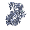
- Structure visualization
| Structure viewer | Molecule: MolmilJmol/JSmol |
|---|
- Downloads & links
-Download
| PDBx/mmCIF format | 7cnt.cif.gz | 413.6 KB | Display | PDBx/mmCIF format |
|---|---|---|---|---|
| PDB format | pdb7cnt.ent.gz | 342.1 KB | Display | PDB format |
| PDBx/mmJSON format | 7cnt.json.gz | Tree view | PDBx/mmJSON format | |
| Others |  Other downloads Other downloads |
-Validation report
| Summary document | 7cnt_validation.pdf.gz | 2.4 MB | Display | wwPDB validaton report |
|---|---|---|---|---|
| Full document | 7cnt_full_validation.pdf.gz | 2.4 MB | Display | |
| Data in XML | 7cnt_validation.xml.gz | 72.4 KB | Display | |
| Data in CIF | 7cnt_validation.cif.gz | 99.7 KB | Display | |
| Arichive directory | https://data.pdbj.org/pub/pdb/validation_reports/cn/7cntftp://data.pdbj.org/pub/pdb/validation_reports/cn/7cnt | HTTPS FTP |
-Related structure data


-Links
 PDBj
PDBj


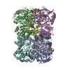
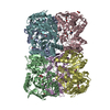
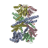
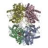
- Assembly
| Deposited unit | 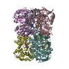
| ||||||||
|---|---|---|---|---|---|---|---|---|---|
| 1 |
| ||||||||
| Unit cell |
|
-Components
| #1: Protein | Mass: 41152.680 Da / Num. of mol.: 6 Source method: isolated from a genetically manipulated source Source: (gene. exp.) Pseudomonas putida KT2440 (bacteria) / Strain: KT2440 / Gene: nicX, PP_3945 / Production host: References: UniProt: Q88FY1, 2,5-dihydroxypyridine 5,6-dioxygenase #2: Chemical | ChemComp-FE2 /   Mass: 55.845 Da / Num. of mol.: 6 / Source method: obtained synthetically / Formula: Fe / Feature type: SUBJECT OF INVESTIGATION Mass: 55.845 Da / Num. of mol.: 6 / Source method: obtained synthetically / Formula: Fe / Feature type: SUBJECT OF INVESTIGATION#3: Water | ChemComp-HOH / |  Mass: 18.015 Da / Num. of mol.: 241 / Source method: isolated from a natural source / Formula: H2O Mass: 18.015 Da / Num. of mol.: 241 / Source method: isolated from a natural source / Formula: H2OHas ligand of interest | Y | Has protein modification | Y | |
|---|
-Experimental details
-Experiment
| Experiment | Method: X-RAY DIFFRACTION / Number of used crystals: 1 |
|---|
- Sample preparation
| Crystal | Density Matthews: 2.33 Å3/Da / Density % sol: 47.17 % |
|---|---|
| Crystal grow | Temperature: 293 K / Method: vapor diffusion, hanging drop / Details: 0.2M Succnic acid PH7 20%PEG3350 |
-Data collection
| Diffraction | Mean temperature: 100 K / Serial crystal experiment: N |
|---|---|
| Diffraction source | Source: SYNCHROTRON / Site: SSRF / Beamline: BL17U / Wavelength: 0.9789 Å |
| Detector | Type: ADSC QUANTUM 315r / Detector: CCD / Date: Mar 28, 2018 |
| Radiation | Protocol: SINGLE WAVELENGTH / Monochromatic (M) / Laue (L): M / Scattering type: x-ray |
| Radiation wavelength | Wavelength: 0.9789 Å / Relative weight: 1 |
| Reflection | Resolution: 2.28→50 Å / Num. obs: 98134 / % possible obs: 99.2 % / Redundancy: 6 % / Rrim(I) all: 0.101 / Net I/σ(I): 8.7 |
| Reflection shell | Resolution: 2.28→2.4 Å / Mean I/σ(I) obs: 3 / Num. unique obs: 14230 / Rrim(I) all: 0.711 |
- Processing
| Software |
| ||||||||||||||||||||||||||||||||||||||||||||||||||||||||||||
|---|---|---|---|---|---|---|---|---|---|---|---|---|---|---|---|---|---|---|---|---|---|---|---|---|---|---|---|---|---|---|---|---|---|---|---|---|---|---|---|---|---|---|---|---|---|---|---|---|---|---|---|---|---|---|---|---|---|---|---|---|---|
| Refinement | Method to determine structure: SAD / Resolution: 2.28→30 Å / Cor.coef. Fo:Fc: 0.967 / Cor.coef. Fo:Fc free: 0.945 / Cross valid method: THROUGHOUT / σ(F): 0 / ESU R: 0.387 / ESU R Free: 0.263 / Stereochemistry target values: MAXIMUM LIKELIHOOD Details: HYDROGENS HAVE BEEN ADDED IN THE RIDING POSITIONS U VALUES : REFINED INDIVIDUALLY
| ||||||||||||||||||||||||||||||||||||||||||||||||||||||||||||
| Solvent computation | Ion probe radii: 0.8 Å / Shrinkage radii: 0.8 Å / VDW probe radii: 1.2 Å / Solvent model: BABINET MODEL WITH MASK | ||||||||||||||||||||||||||||||||||||||||||||||||||||||||||||
| Displacement parameters | Biso max: 173.03 Å2 / Biso mean: 67.306 Å2 / Biso min: 32.03 Å2
| ||||||||||||||||||||||||||||||||||||||||||||||||||||||||||||
| Refinement step | Cycle: final / Resolution: 2.28→30 Å
| ||||||||||||||||||||||||||||||||||||||||||||||||||||||||||||
| Refine LS restraints |
| ||||||||||||||||||||||||||||||||||||||||||||||||||||||||||||
| LS refinement shell | Resolution: 2.28→2.339 Å / Rfactor Rfree error: 0 / Total num. of bins used: 20
|
