 Movie
Movie Controller

[English] 日本語
 Yorodumi
- PDB-7cne: Crystal Structure of Sphingomyelinase C from Streptomyces griseoc... -
+ Open data

- Basic information
| Entry | Database: PDB / ID: 7cne | |||||||||
|---|---|---|---|---|---|---|---|---|---|---|
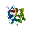
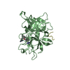
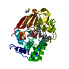
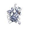
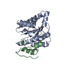
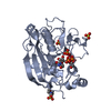
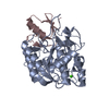
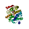
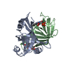
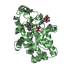
| Title | Crystal Structure of Sphingomyelinase C from Streptomyces griseocarneus | |||||||||
 Components Components | Sphingomyelinase C | |||||||||
 Keywords Keywords | HYDROLASE / Sphingomyelinase C / Streptomyces griseocarneus / Metal-binding site | |||||||||
| Function / homology |  Function and homology information Function and homology informationsphingomyelin phosphodiesterase / sphingomyelin phosphodiesterase activity / extracellular region Similarity search - Function | |||||||||
| Biological species |  Streptomyces griseocarneus (bacteria) Streptomyces griseocarneus (bacteria) | |||||||||
| Method |  X-RAY DIFFRACTION / MOLECULAR REPLACEMENT / Resolution: 2 Å X-RAY DIFFRACTION / MOLECULAR REPLACEMENT / Resolution: 2 Å | |||||||||
 Authors Authors | Murayama, K. / Fujisawa, I. / Sugimori, D. | |||||||||
 Citation Citation | Journal: J.Biosci.Bioeng. / Year: 2021 Title: Structural basis for the high thermal stability and optimum pH of sphingomyelinase C from Streptomyces griseocarneus. Authors: Fujisawa, I. / Hamana, H. / Tomita, Y. / Matsumoto, Y. / Murayama, K. / Sugimori, D. | |||||||||
| History |
|
- Structure visualization
| Structure viewer | Molecule: MolmilJmol/JSmol |
|---|
- Downloads & links
-Download
| PDBx/mmCIF format | 7cne.cif.gz | 94.3 KB | Display | PDBx/mmCIF format |
|---|---|---|---|---|
| PDB format | pdb7cne.ent.gz | 56 KB | Display | PDB format |
| PDBx/mmJSON format | 7cne.json.gz | Tree view | PDBx/mmJSON format | |
| Others |  Other downloads Other downloads |
-Validation report
| Arichive directory | https://data.pdbj.org/pub/pdb/validation_reports/cn/7cneftp://data.pdbj.org/pub/pdb/validation_reports/cn/7cne | HTTPS FTP |
|---|
-Related structure data
| Related structure data | 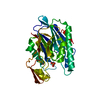 1zwxS S: Starting model for refinement |
|---|---|
| Similar structure data |
-Links
 PDBj
PDBj

- Assembly
| Deposited unit | 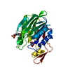
| ||||||||||||
|---|---|---|---|---|---|---|---|---|---|---|---|---|---|
| 1 |
| ||||||||||||
| Unit cell |
|
-Components
| #1: Protein | Mass: 32216.322 Da / Num. of mol.: 1 Source method: isolated from a genetically manipulated source Source: (gene. exp.) Streptomyces griseocarneus (bacteria) / Gene: smc / Production host: Streptomyces lividans (bacteria)References: UniProt: A6P7M9, sphingomyelin phosphodiesterase |
|---|---|
| #2: Chemical | ChemComp-EPE /   Mass: 238.305 Da / Num. of mol.: 1 / Source method: obtained synthetically / Formula: C8H18N2O4S / Comment: pH buffer*YM Mass: 238.305 Da / Num. of mol.: 1 / Source method: obtained synthetically / Formula: C8H18N2O4S / Comment: pH buffer*YM |
| #3: Chemical | ChemComp-SIN /   Mass: 118.088 Da / Num. of mol.: 1 / Source method: obtained synthetically / Formula: C4H6O4 Mass: 118.088 Da / Num. of mol.: 1 / Source method: obtained synthetically / Formula: C4H6O4 |
| #4: Water | ChemComp-HOH /  Mass: 18.015 Da / Num. of mol.: 303 / Source method: isolated from a natural source / Formula: H2O Mass: 18.015 Da / Num. of mol.: 303 / Source method: isolated from a natural source / Formula: H2O |
| Has ligand of interest | N |
| Has protein modification | Y |
-Experimental details
-Experiment
| Experiment | Method: X-RAY DIFFRACTION / Number of used crystals: 1 |
|---|
- Sample preparation
| Crystal | Density Matthews: 2.41 Å3/Da / Density % sol: 49.05 % |
|---|---|
| Crystal grow | Temperature: 298 K / Method: vapor diffusion, hanging drop Details: 0.95M succinic acid, 1% PEGMME 2000, 0.1M HEPES buffer pH 7.0 |
-Data collection
| Diffraction | Mean temperature: 100 K / Serial crystal experiment: N |
|---|---|
| Diffraction source | Source: ROTATING ANODE / Type: RIGAKU FR-E SUPERBRIGHT / Wavelength: 1.5418 Å |
| Detector | Type: RIGAKU / Detector: IMAGE PLATE / Date: Jul 11, 2007 |
| Radiation | Protocol: SINGLE WAVELENGTH / Monochromatic (M) / Laue (L): M / Scattering type: x-ray |
| Radiation wavelength | Wavelength: 1.5418 Å / Relative weight: 1 |
| Reflection | Resolution: 2→48.5 Å / Num. obs: 22156 / % possible obs: 99.8 % / Redundancy: 13.4 % / Biso Wilson estimate: 24.02 Å2 / Rmerge(I) obs: 0.105 / Net I/σ(I): 36.6 |
| Reflection shell | Resolution: 2→2.07 Å / Num. unique obs: 2162 / CC1/2: 0.979 |
- Processing
| Software |
| |||||||||||||||||||||||||||||||||||||||||||||||||||||||||||||||||||||||||||||||||||||||||||||||||||||||||
|---|---|---|---|---|---|---|---|---|---|---|---|---|---|---|---|---|---|---|---|---|---|---|---|---|---|---|---|---|---|---|---|---|---|---|---|---|---|---|---|---|---|---|---|---|---|---|---|---|---|---|---|---|---|---|---|---|---|---|---|---|---|---|---|---|---|---|---|---|---|---|---|---|---|---|---|---|---|---|---|---|---|---|---|---|---|---|---|---|---|---|---|---|---|---|---|---|---|---|---|---|---|---|---|---|---|---|
| Refinement | Method to determine structure: MOLECULAR REPLACEMENT Starting model: 1ZWX Resolution: 2→48.45 Å / SU ML: 0.1672 / Cross valid method: FREE R-VALUE / σ(F): 1.34 / Phase error: 20.0148 / Stereochemistry target values: GeoStd + Monomer Library
| |||||||||||||||||||||||||||||||||||||||||||||||||||||||||||||||||||||||||||||||||||||||||||||||||||||||||
| Solvent computation | Shrinkage radii: 0.9 Å / VDW probe radii: 1.11 Å / Solvent model: FLAT BULK SOLVENT MODEL | |||||||||||||||||||||||||||||||||||||||||||||||||||||||||||||||||||||||||||||||||||||||||||||||||||||||||
| Displacement parameters | Biso mean: 24.51 Å2 | |||||||||||||||||||||||||||||||||||||||||||||||||||||||||||||||||||||||||||||||||||||||||||||||||||||||||
| Refinement step | Cycle: LAST / Resolution: 2→48.45 Å
| |||||||||||||||||||||||||||||||||||||||||||||||||||||||||||||||||||||||||||||||||||||||||||||||||||||||||
| Refine LS restraints |
| |||||||||||||||||||||||||||||||||||||||||||||||||||||||||||||||||||||||||||||||||||||||||||||||||||||||||
| LS refinement shell |
|
