 Movie
Movie Controller

[English] 日本語
 Yorodumi
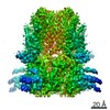
- PDB-7cn1: Cryo-EM structure of K+-bound hERG channel in the presence of ast... -
+ Open data

- Basic information
| Entry | Database: PDB / ID: 7cn1 | ||||||||||||
|---|---|---|---|---|---|---|---|---|---|---|---|---|---|
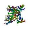
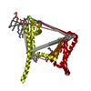
| Title | Cryo-EM structure of K+-bound hERG channel in the presence of astemizole | ||||||||||||
 Components Components | potassium channel | ||||||||||||
 Keywords Keywords | TRANSPORT PROTEIN / potassium channel | ||||||||||||
| Function / homology | :  Function and homology information Function and homology information | ||||||||||||
| Biological species |  Homo sapiens (human) Homo sapiens (human) | ||||||||||||
| Method | ELECTRON MICROSCOPY / single particle reconstruction / cryo EM / Resolution: 3.7 Å | ||||||||||||
 Authors Authors | Asai, T. / Adachi, N. / Moriya, T. / Kawasaki, M. / Suzuki, K. / Senda, T. / Murata, T. | ||||||||||||
| Funding support |  Japan, 3items Japan, 3items
| ||||||||||||
 Citation Citation | Journal: Structure / Year: 2021 Title: Cryo-EM Structure of K-Bound hERG Channel Complexed with the Blocker Astemizole. Authors: Tatsuki Asai / Naruhiko Adachi / Toshio Moriya / Hideyuki Oki / Takamitsu Maru / Masato Kawasaki / Kano Suzuki / Sisi Chen / Ryohei Ishii / Kazuko Yonemori / Shigeru Igaki / Satoshi Yasuda / ...Authors: Tatsuki Asai / Naruhiko Adachi / Toshio Moriya / Hideyuki Oki / Takamitsu Maru / Masato Kawasaki / Kano Suzuki / Sisi Chen / Ryohei Ishii / Kazuko Yonemori / Shigeru Igaki / Satoshi Yasuda / Satoshi Ogasawara / Toshiya Senda / Takeshi Murata / Abstract: The hERG channel is a voltage-gated potassium channel involved in cardiac repolarization. Off-target hERG inhibition by drugs has become a critical issue in the pharmaceutical industry. The three- ...The hERG channel is a voltage-gated potassium channel involved in cardiac repolarization. Off-target hERG inhibition by drugs has become a critical issue in the pharmaceutical industry. The three-dimensional structure of the hERG channel was recently reported at 3.8-Å resolution using cryogenic electron microscopy (cryo-EM). However, the drug inhibition mechanism remains unclear because of the scarce structural information regarding the drug- and potassium-bound hERG channels. In this study, we obtained the cryo-EM density map of potassium-bound hERG channel complexed with astemizole, a well-known hERG inhibitor that increases risk of potentially fatal arrhythmia, at 3.5-Å resolution. The structure suggested that astemizole inhibits potassium conduction by binding directly below the selectivity filter. Furthermore, we propose a possible binding model of astemizole to the hERG channel and provide insights into the unusual sensitivity of hERG to several drugs. | ||||||||||||
| History |
|
- Structure visualization
| Movie |
Movie viewer |
|---|---|
| Structure viewer | Molecule: MolmilJmol/JSmol |
- Downloads & links
-Download
| PDBx/mmCIF format | 7cn1.cif.gz | 196.8 KB | Display | PDBx/mmCIF format |
|---|---|---|---|---|
| PDB format | pdb7cn1.ent.gz | 127.8 KB | Display | PDB format |
| PDBx/mmJSON format | 7cn1.json.gz | Tree view | PDBx/mmJSON format | |
| Others |  Other downloads Other downloads |
-Validation report
| Arichive directory | https://data.pdbj.org/pub/pdb/validation_reports/cn/7cn1ftp://data.pdbj.org/pub/pdb/validation_reports/cn/7cn1 | HTTPS FTP |
|---|
-Related structure data
| Related structure data |  30413MC  7cn0C M: map data used to model this data C: citing same article ( |
|---|---|
| Similar structure data | |
| EM raw data | EMPIAR-10629 (Title: Cryo-EM structure of K+-bound hERG channel in the presence of astemizole Data size: 1.8 TB Data #1: Cryo-EM structure of K+-bound hERG channel in the presence of astemizole [micrographs - multiframe]) |


-Links
 PDBj
PDBj
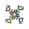
- Assembly
| Deposited unit | 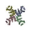
|
|---|---|
| 1 |
|
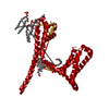
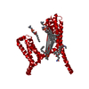
-Components
| #1: Protein | Mass: 91792.086 Da / Num. of mol.: 4 Source method: isolated from a genetically manipulated source Source: (gene. exp.) Homo sapiens (human) / Plasmid: BacMam expression vector / Production host: Homo sapiens (human) / Strain (production host): Expi293F cells#2: Chemical |   Mass: 39.098 Da / Num. of mol.: 3 / Source method: obtained synthetically / Formula: K / Feature type: SUBJECT OF INVESTIGATION Mass: 39.098 Da / Num. of mol.: 3 / Source method: obtained synthetically / Formula: K / Feature type: SUBJECT OF INVESTIGATIONHas ligand of interest | Y | |
|---|
-Experimental details
-Experiment
| Experiment | Method: ELECTRON MICROSCOPY |
|---|---|
| EM experiment | Aggregation state: PARTICLE / 3D reconstruction method: single particle reconstruction |
- Sample preparation
| Component | Name: potassium channel / Type: ORGANELLE OR CELLULAR COMPONENT / Details: homo-tetramer / Entity ID: #1 / Source: RECOMBINANT |
|---|---|
| Source (natural) | Organism: Homo sapiens (human) |
| Source (recombinant) | Organism: Homo sapiens (human) / Cell: Expi293F cells / Plasmid: BacMam expression vector |
| Buffer solution | pH: 7.4 |
| Specimen | Conc.: 2 mg/ml / Embedding applied: NO / Shadowing applied: NO / Staining applied: NO / Vitrification applied: YES / Details: This sample was mono-disperse. |
| Specimen support | Details: The grid was washed by acetone prior to use. / Grid material: COPPER / Grid mesh size: 300 divisions/in. / Grid type: Quantifoil R1.2/1.3 |
| Vitrification | Instrument: FEI VITROBOT MARK IV / Cryogen name: ETHANE / Humidity: 100 % / Chamber temperature: 291 K / Details: Blotting time was 5 seconds (blot force 10) |
- Electron microscopy imaging
| Experimental equipment |  Model: Talos Arctica / Image courtesy: FEI Company |
|---|---|
| Microscopy | Model: FEI TALOS ARCTICA |
| Electron gun | Electron source:  FIELD EMISSION GUN / Accelerating voltage: 200 kV / Illumination mode: FLOOD BEAM FIELD EMISSION GUN / Accelerating voltage: 200 kV / Illumination mode: FLOOD BEAM |
| Electron lens | Mode: BRIGHT FIELD / Nominal magnification: 120000 X / Nominal defocus max: 2000 nm / Nominal defocus min: 800 nm / Cs: 2.7 mm / C2 aperture diameter: 50 µm |
| Specimen holder | Cryogen: NITROGEN |
| Image recording | Average exposure time: 55.55 sec. / Electron dose: 50 e/Å2 / Detector mode: COUNTING / Film or detector model: FEI FALCON III (4k x 4k) / Num. of grids imaged: 1 / Num. of real images: 1865 |
- Processing
| Software | Name: PHENIX / Version: 1.16_3549: / Classification: refinement | ||||||||||||||||||||||||||||||||||||||||
|---|---|---|---|---|---|---|---|---|---|---|---|---|---|---|---|---|---|---|---|---|---|---|---|---|---|---|---|---|---|---|---|---|---|---|---|---|---|---|---|---|---|
| EM software |
| ||||||||||||||||||||||||||||||||||||||||
| CTF correction | Type: PHASE FLIPPING AND AMPLITUDE CORRECTION | ||||||||||||||||||||||||||||||||||||||||
| Particle selection | Num. of particles selected: 930193 | ||||||||||||||||||||||||||||||||||||||||
| Symmetry | Point symmetry: C1 (asymmetric) | ||||||||||||||||||||||||||||||||||||||||
| 3D reconstruction | Resolution: 3.7 Å / Resolution method: FSC 0.143 CUT-OFF / Num. of particles: 102128 / Symmetry type: POINT | ||||||||||||||||||||||||||||||||||||||||
| Atomic model building | Protocol: AB INITIO MODEL / Space: REAL | ||||||||||||||||||||||||||||||||||||||||
| Refine LS restraints |
|
