| 登録情報 | データベース: PDB / ID: 7clu
|
|---|
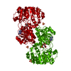
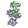
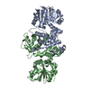
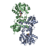
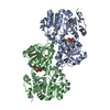
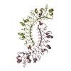
| タイトル | PigF with SAH |
|---|
 要素 要素 | Methyltransferase domain-containing protein |
|---|
 キーワード キーワード | TRANSFERASE / prodigiosin / PigF / methyl-transferase / induce-fit |
|---|
| 機能・相同性 |  機能・相同性情報 機能・相同性情報
O-methyltransferase activity / methylation / protein dimerization activity類似検索 - 分子機能 Plant methyltransferase dimerisation / O-methyltransferase dimerisation domain / O-methyltransferase domain / O-methyltransferase COMT-type / O-methyltransferase domain / SAM-dependent O-methyltransferase class II-type profile. / Vaccinia Virus protein VP39 / Winged helix-like DNA-binding domain superfamily/Winged helix DNA-binding domain / Arc Repressor Mutant, subunit A / Winged helix DNA-binding domain superfamily ...Plant methyltransferase dimerisation / O-methyltransferase dimerisation domain / O-methyltransferase domain / O-methyltransferase COMT-type / O-methyltransferase domain / SAM-dependent O-methyltransferase class II-type profile. / Vaccinia Virus protein VP39 / Winged helix-like DNA-binding domain superfamily/Winged helix DNA-binding domain / Arc Repressor Mutant, subunit A / Winged helix DNA-binding domain superfamily / Winged helix-like DNA-binding domain superfamily / S-adenosyl-L-methionine-dependent methyltransferase superfamily / Rossmann fold / Orthogonal Bundle / 3-Layer(aba) Sandwich / Mainly Alpha / Alpha Beta類似検索 - ドメイン・相同性 ACETATE ION / Methyltransferase domain-containing protein類似検索 - 構成要素 |
|---|
| 生物種 |  Serratia marcescens (霊菌) Serratia marcescens (霊菌) |
|---|
| 手法 |  X線回折 / シンクロトロン / 単波長異常分散 / 解像度: 1.9 Å X線回折 / シンクロトロン / 単波長異常分散 / 解像度: 1.9 Å |
|---|
 データ登録者 データ登録者 | Qiu, S. / Xu, D. / Han, N. / Sun, B. / Ran, T. / Wang, W. |
|---|
| 資金援助 |  中国, 2件 中国, 2件 | 組織 | 認可番号 | 国 |
|---|
| National Natural Science Foundation of China (NSFC) | 31770074 | 中国 | | National Natural Science Foundation of China (NSFC) | 31770050 | 中国 |
|
|---|
 引用 引用 | ジャーナル: Iucrj / 年: 2022タイトル: Crystal structures of PigF, an O-methyltransferase involved in the prodigiosin synthetic pathway, reveal an induced-fit substrate-recognition mechanism. 著者: Qiu, S. / Xu, D. / Xu, M. / Zhou, H. / Sun, N. / Zhang, L. / Zhao, M. / He, J. / Ran, T. / Sun, B. / Wang, W. |
|---|
| 履歴 | | 登録 | 2020年7月22日 | 登録サイト: PDBJ / 処理サイト: PDBJ |
|---|
| 改定 1.0 | 2021年7月28日 | Provider: repository / タイプ: Initial release |
|---|
| 改定 1.1 | 2022年3月16日 | Group: Database references / カテゴリ: citation / citation_author / database_2
Item: _citation.country / _citation.journal_abbrev ..._citation.country / _citation.journal_abbrev / _citation.journal_id_CSD / _citation.journal_id_ISSN / _citation.journal_volume / _citation.page_first / _citation.page_last / _citation.pdbx_database_id_DOI / _citation.title / _citation.year / _database_2.pdbx_DOI / _database_2.pdbx_database_accession |
|---|
| 改定 1.2 | 2024年5月29日 | Group: Data collection / Derived calculations / カテゴリ: atom_type / chem_comp_atom / chem_comp_bond / Item: _atom_type.pdbx_N_electrons / _atom_type.pdbx_scat_Z |
|---|
|
|---|
 ムービー
ムービー コントローラー


 データを開く
 基本情報
基本情報 構造の表示
構造の表示 ダウンロードとリンク
 その他のダウンロード
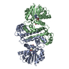


 PDBj
PDBj 集合体
集合体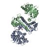

 分子量: 59.044 Da / 分子数: 1 / 由来タイプ: 合成 / 式: C2H3O2 / タイプ: SUBJECT OF INVESTIGATION
 分子量: 92.094 Da / 分子数: 1 / 由来タイプ: 合成 / 式: C3H8O3 / タイプ: SUBJECT OF INVESTIGATION
 分子量: 18.015 Da / 分子数: 334 / 由来タイプ: 天然 / 式: H2O
 試料調製
試料調製 解析
解析