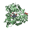
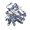
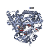
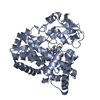
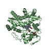
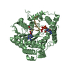
[English] 日本語
Yorodumi
- PDB-7cl9: Androstenedione-bound structure of CYP154C2 from Streptomyces ave... -

+
Open data


ID or keywords:

Loading...

-
Basic information

Entry
Database: PDB / ID: 7cl9
TitleAndrostenedione-bound structure of CYP154C2 from Streptomyces avermitilis in an open conformation
ComponentsCytochrome P450 hydroxylase
KeywordsOXIDOREDUCTASE / Cytochrome P450 / hydroxylase / Androstenedione
Function / homology
Function and homology information


cholest-4-en-3-one 26-monooxygenase activity / steroid hydroxylase activity / cholesterol catabolic process / iron ion binding / heme binding
Similarity search - Function
Cytochrome P450, B-class / Cytochrome P450, conserved site / Cytochrome P450 cysteine heme-iron ligand signature. / Cytochrome P450 / Cytochrome P450 superfamily / Cytochrome P450
Similarity search - Domain/homology
4-ANDROSTENE-3-17-DIONE / PROTOPORPHYRIN IX CONTAINING FE / DI(HYDROXYETHYL)ETHER / Cytochrome P450 hydroxylase
Similarity search - Component
Biological speciesStreptomyces avermitilis MA-4680 = NBRC 14893 (bacteria)
MethodX-RAY DIFFRACTION / SYNCHROTRON / MOLECULAR REPLACEMENT / Resolution: 1.95 Å
AuthorsXu, L.H. / Fushinobu, S.
Funding support China, 1items
OrganizationGrant numberCountry
National Natural Science Foundation of China (NSFC)81402810 China
CitationJournal: To Be Published
Title: Androstenedione-bound structure of CYP154C2 from Streptomyces avermitilis in an open conformation
Authors: Xu, L.H. / Fushinobu, S.
History
DepositionJul 20, 2020Deposition site: PDBJ / Processing site: PDBJ
Revision 1.0Jul 21, 2021Provider: repository / Type: Initial release
Revision 2.0Sep 20, 2023Group: Atomic model / Author supporting evidence ...Atomic model / Author supporting evidence / Data collection / Database references / Derived calculations / Non-polymer description / Refinement description / Source and taxonomy / Structure summary
Category: atom_site / chem_comp ...atom_site / chem_comp / chem_comp_atom / chem_comp_bond / database_2 / diffrn_detector / entity / entity_src_gen / pdbx_contact_author / pdbx_entity_instance_feature / pdbx_entity_nonpoly / pdbx_entry_details / pdbx_nonpoly_scheme / pdbx_struct_assembly_gen / pdbx_struct_conn_angle / refine / refine_hist / refine_ls_restr / refine_ls_shell / struct_asym / struct_conn
Item: _chem_comp.formula / _chem_comp.formula_weight ..._chem_comp.formula / _chem_comp.formula_weight / _chem_comp.id / _chem_comp.mon_nstd_flag / _chem_comp.name / _chem_comp.pdbx_synonyms / _chem_comp.type / _database_2.pdbx_DOI / _database_2.pdbx_database_accession / _diffrn_detector.detector / _diffrn_detector.type / _entity_src_gen.pdbx_gene_src_scientific_name / _pdbx_entry_details.has_ligand_of_interest / _pdbx_struct_assembly_gen.asym_id_list / _pdbx_struct_conn_angle.ptnr3_label_asym_id / _refine.B_iso_max / _refine.B_iso_min / _refine.details / _refine.ls_R_factor_R_work / _refine.ls_R_factor_obs / _refine.pdbx_ls_sigma_F / _refine_hist.cycle_id / _refine_hist.number_atoms_total / _refine_hist.pdbx_B_iso_mean_ligand / _refine_hist.pdbx_B_iso_mean_solvent / _refine_hist.pdbx_number_atoms_ligand / _refine_hist.pdbx_number_residues_total / _refine_ls_shell.R_factor_R_free_error / _refine_ls_shell.number_reflns_all / _struct_conn.ptnr2_label_asym_id
Description: Atoms with unrealistic or zero occupancies / Provider: author / Type: Coordinate replacement
Revision 2.1Nov 29, 2023Group: Refinement description / Category: pdbx_initial_refinement_model

-
Structure visualization

Structure viewerMolecule:
MolmilJmol/JSmol

Downloads & links

-
Assembly

Deposited unit
A: Cytochrome P450 hydroxylase
hetero molecules


Theoretical massNumber of molelcules
Total (without water)44,1104
Polymers43,1011
Non-polymers1,0093
Water5,477304
1


  • Idetical with deposited unit
  • defined by author&software
TypeNameSymmetry operationNumber
identity operation1_555x,y,z1
Buried area1190 Å2
ΔGint-24 kcal/mol
Surface area17600 Å2
MethodPISA
Unit cell
Length a, b, c (Å)54.600, 80.130, 120.630
Angle α, β, γ (deg.)90.00, 90.00, 90.00
Int Tables number19
Space group name H-MP212121

-
Components

#1: Protein Cytochrome P450 hydroxylase / CYP154C2


Mass: 43101.059 Da / Num. of mol.: 1
Source method: isolated from a genetically manipulated source
Source: (gene. exp.) Streptomyces avermitilis MA-4680 = NBRC 14893 (bacteria)
Strain: ATCC 31267 / DSM 46492 / JCM 5070 / NBRC 14893 / NCIMB 12804 / NRRL 8165 / MA-4680
Gene: cyp19, SAVERM_3882 / Production host: Escherichia coli (E. coli) / References: UniProt: Q82GL5
#2: Chemical ChemComp-HEM / PROTOPORPHYRIN IX CONTAINING FE / HEME


Mass: 616.487 Da / Num. of mol.: 1 / Source method: obtained synthetically / Formula: C34H32FeN4O4
#3: Chemical ChemComp-ASD / 4-ANDROSTENE-3-17-DIONE


Mass: 286.409 Da / Num. of mol.: 1 / Source method: obtained synthetically / Formula: C19H26O2 / Comment: hormone*YM
#4: Chemical ChemComp-PEG / DI(HYDROXYETHYL)ETHER


Mass: 106.120 Da / Num. of mol.: 1 / Source method: obtained synthetically / Formula: C4H10O3
#5: Water ChemComp-HOH / water


Mass: 18.015 Da / Num. of mol.: 304 / Source method: isolated from a natural source / Formula: H2O
Has ligand of interestN

-
Experimental details

-
Experiment

ExperimentMethod: X-RAY DIFFRACTION / Number of used crystals: 1

-
Sample preparation

CrystalDensity Matthews: 3.06 Å3/Da / Density % sol: 59.82 %
Crystal growTemperature: 293 K / Method: vapor diffusion, sitting drop / pH: 7.5
Details: 0.06 M Divalents, 0.1 M Buffer System 2 pH 7.5, 50% v/v Precipitant mix 1

-
Data collection

DiffractionMean temperature: 298 K / Serial crystal experiment: N
Diffraction sourceSource: SYNCHROTRON / Site: SSRF / Beamline: BL18U1 / Wavelength: 0.979 Å
DetectorType: DECTRIS PILATUS 6M / Detector: PIXEL / Date: Mar 16, 2019
RadiationProtocol: SINGLE WAVELENGTH / Monochromatic (M) / Laue (L): M / Scattering type: x-ray
Radiation wavelengthWavelength: 0.979 Å / Relative weight: 1
ReflectionResolution: 1.95→50 Å / Num. obs: 39415 / % possible obs: 100 % / Redundancy: 11.6 % / CC1/2: 0.998 / Rmerge(I) obs: 0.098 / Net I/σ(I): 13.5
Reflection shellResolution: 1.95→2.06 Å / Redundancy: 11.5 % / Rmerge(I) obs: 0.932 / Mean I/σ(I) obs: 2.8 / Num. unique obs: 5656 / CC1/2: 0.851 / % possible all: 100

-
Processing

Software
NameVersionClassification
REFMAC5.8.0238refinement
PDB_EXTRACT3.25data extraction
iMOSFLMdata reduction
SCALAdata scaling
MOLREPphasing
RefinementMethod to determine structure: MOLECULAR REPLACEMENT
Starting model: 6L69
Resolution: 1.95→49.79 Å / Cor.coef. Fo:Fc: 0.963 / Cor.coef. Fo:Fc free: 0.927 / SU B: 4.178 / SU ML: 0.113 / Cross valid method: THROUGHOUT / ESU R: 0.144 / ESU R Free: 0.149 / Stereochemistry target values: MAXIMUM LIKELIHOOD / Details: HYDROGENS HAVE BEEN ADDED IN THE RIDING POSITIONS
RfactorNum. reflection% reflectionSelection details
Rfree0.2488 2001 5.1 %RANDOM
Rwork0.19208 ---
obs0.19495 37351 99.99 %-
Solvent computationIon probe radii: 0.8 Å / Shrinkage radii: 0.8 Å / VDW probe radii: 1.2 Å / Solvent model: MASK
Displacement parametersBiso mean: 40.571 Å2
Baniso -1Baniso -2Baniso -3
1--0 Å2-0 Å2-0 Å2
2---0 Å20 Å2
3---0 Å2
Refinement stepCycle: 1 / Resolution: 1.95→49.79 Å
ProteinNucleic acidLigandSolventTotal
Num. atoms3017 0 71 304 3392
Refine LS restraints
Refine-IDTypeDev idealDev ideal targetNumber
X-RAY DIFFRACTIONr_bond_refined_d0.010.0133159
X-RAY DIFFRACTIONr_bond_other_d0.0010.0173014
X-RAY DIFFRACTIONr_angle_refined_deg1.621.6664333
X-RAY DIFFRACTIONr_angle_other_deg1.3691.586954
X-RAY DIFFRACTIONr_dihedral_angle_1_deg6.4285397
X-RAY DIFFRACTIONr_dihedral_angle_2_deg32.56321.118152
X-RAY DIFFRACTIONr_dihedral_angle_3_deg15.04615490
X-RAY DIFFRACTIONr_dihedral_angle_4_deg20.9151526
X-RAY DIFFRACTIONr_chiral_restr0.0770.2420
X-RAY DIFFRACTIONr_gen_planes_refined0.0090.023551
X-RAY DIFFRACTIONr_gen_planes_other0.010.02643
X-RAY DIFFRACTIONr_nbd_refined
X-RAY DIFFRACTIONr_nbd_other
X-RAY DIFFRACTIONr_nbtor_refined
X-RAY DIFFRACTIONr_nbtor_other
X-RAY DIFFRACTIONr_xyhbond_nbd_refined
X-RAY DIFFRACTIONr_xyhbond_nbd_other
X-RAY DIFFRACTIONr_metal_ion_refined
X-RAY DIFFRACTIONr_metal_ion_other
X-RAY DIFFRACTIONr_symmetry_vdw_refined
X-RAY DIFFRACTIONr_symmetry_vdw_other
X-RAY DIFFRACTIONr_symmetry_hbond_refined
X-RAY DIFFRACTIONr_symmetry_hbond_other
X-RAY DIFFRACTIONr_symmetry_metal_ion_refined
X-RAY DIFFRACTIONr_symmetry_metal_ion_other
X-RAY DIFFRACTIONr_mcbond_it3.6984.1191591
X-RAY DIFFRACTIONr_mcbond_other3.6964.1151589
X-RAY DIFFRACTIONr_mcangle_it5.0776.1721987
X-RAY DIFFRACTIONr_mcangle_other5.0766.1711987
X-RAY DIFFRACTIONr_scbond_it4.3264.4951568
X-RAY DIFFRACTIONr_scbond_other4.3244.4951569
X-RAY DIFFRACTIONr_scangle_it
X-RAY DIFFRACTIONr_scangle_other6.3436.5982347
X-RAY DIFFRACTIONr_long_range_B_refined8.15150.1343647
X-RAY DIFFRACTIONr_long_range_B_other8.15449.7523568
X-RAY DIFFRACTIONr_rigid_bond_restr
X-RAY DIFFRACTIONr_sphericity_free
X-RAY DIFFRACTIONr_sphericity_bonded
LS refinement shellResolution: 1.95→2.001 Å / Total num. of bins used: 20
RfactorNum. reflection% reflection
Rfree0.292 140 -
Rwork0.31 2707 -
obs--100 %

+
About Yorodumi

-
News

-
Feb 9, 2022. New format data for meta-information of EMDB entries

New format data for meta-information of EMDB entries

  • Version 3 of the EMDB header file is now the official format.
  • The previous official version 1.9 will be removed from the archive.

Related info.:EMDB header

External links:wwPDB to switch to version 3 of the EMDB data model

-
Aug 12, 2020. Covid-19 info

Covid-19 info

URL: https://pdbjlvh1.pdbj.org/emnavi/covid19.php

New page: Covid-19 featured information page in EM Navigator.

Related info.:Covid-19 info / Mar 5, 2020. Novel coronavirus structure data

+
Mar 5, 2020. Novel coronavirus structure data

Novel coronavirus structure data

Related info.:Yorodumi Speices / Aug 12, 2020. Covid-19 info

External links:COVID-19 featured content - PDBj / Molecule of the Month (242):Coronavirus Proteases

+
Jan 31, 2019. EMDB accession codes are about to change! (news from PDBe EMDB page)

EMDB accession codes are about to change! (news from PDBe EMDB page)

  • The allocation of 4 digits for EMDB accession codes will soon come to an end. Whilst these codes will remain in use, new EMDB accession codes will include an additional digit and will expand incrementally as the available range of codes is exhausted. The current 4-digit format prefixed with “EMD-” (i.e. EMD-XXXX) will advance to a 5-digit format (i.e. EMD-XXXXX), and so on. It is currently estimated that the 4-digit codes will be depleted around Spring 2019, at which point the 5-digit format will come into force.
  • The EM Navigator/Yorodumi systems omit the EMD- prefix.

Related info.:Q: What is EMD? / ID/Accession-code notation in Yorodumi/EM Navigator

External links:EMDB Accession Codes are Changing Soon! / Contact to PDBj

+
Jul 12, 2017. Major update of PDB

Major update of PDB

  • wwPDB released updated PDB data conforming to the new PDBx/mmCIF dictionary.
  • This is a major update changing the version number from 4 to 5, and with Remediation, in which all the entries are updated.
  • In this update, many items about electron microscopy experimental information are reorganized (e.g. em_software).
  • Now, EM Navigator and Yorodumi are based on the updated data.

External links:wwPDB Remediation / Enriched Model Files Conforming to OneDep Data Standards Now Available in the PDB FTP Archive

-
Yorodumi

Thousand views of thousand structures

  • Yorodumi is a browser for structure data from EMDB, PDB, SASBDB, etc.
  • This page is also the successor to EM Navigator detail page, and also detail information page/front-end page for Omokage search.
  • The word "yorodu" (or yorozu) is an old Japanese word meaning "ten thousand". "mi" (miru) is to see.

Related info.:EMDB / PDB / SASBDB / Comparison of 3 databanks / Yorodumi Search / Aug 31, 2016. New EM Navigator & Yorodumi / Yorodumi Papers / Jmol/JSmol / Function and homology information / Changes in new EM Navigator and Yorodumi

Read more