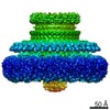
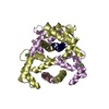
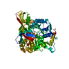
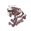
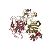
[日本語] English
- PDB-7cik: Structure of the 58-213 fragment of FliF -

+
データを開く


IDまたはキーワード:

読み込み中...

-
基本情報

登録情報
データベース: PDB / ID: 7cik
タイトルStructure of the 58-213 fragment of FliF
要素Flagellar M-ring protein
キーワードMOTOR PROTEIN / Flagellar motor protein
機能・相同性
機能・相同性情報


bacterial-type flagellum basal body, MS ring / cytoskeletal motor activity / bacterial-type flagellum-dependent cell motility / plasma membrane
類似検索 - 分子機能
Flagellar M-ring protein FliF / Flagellar M-ring C-terminal / Flagellar M-ring protein C-terminal / Lipoprotein YscJ/Flagellar M-ring protein / Secretory protein of YscJ/FliF family / Flagellar M-ring , N-terminal / AMP-binding enzyme, C-terminal domain superfamily
類似検索 - ドメイン・相同性
Flagellar M-ring protein
類似検索 - 構成要素
生物種Aquifex aeolicus (バクテリア)
手法X線回折 / シンクロトロン / 単波長異常分散 / 解像度: 2.29 Å
データ登録者Takekawa, T. / Sakuma, M. / Kojima, S. / Homma, M. / Imada, K.
資金援助 日本, 2件
組織認可番号
Japan Society for the Promotion of Science (JSPS)JP16J01859 日本
Japan Society for the Promotion of Science (JSPS)JP15H02386 日本
引用ジャーナル: mBio / : 2021
タイトル: Two Distinct Conformations in 34 FliF Subunits Generate Three Different Symmetries within the Flagellar MS-Ring.
著者: Norihiro Takekawa / Akihiro Kawamoto / Mayuko Sakuma / Takayuki Kato / Seiji Kojima / Miki Kinoshita / Tohru Minamino / Keiichi Namba / Michio Homma / Katsumi Imada /
要旨: The bacterial flagellum is a protein nanomachine essential for bacterial motility. The flagellar basal body contains several ring structures. The MS-ring is embedded in the cytoplasmic membrane and ...The bacterial flagellum is a protein nanomachine essential for bacterial motility. The flagellar basal body contains several ring structures. The MS-ring is embedded in the cytoplasmic membrane and is formed at the earliest stage of flagellar formation to serve as the base for flagellar assembly as well as a housing for the flagellar protein export gate complex. The MS-ring is formed by FliF, which has two transmembrane helices and a large periplasmic region. A recent electron cryomicroscopy (cryoEM) study of the MS-ring formed by overexpressed FliF revealed a symmetry mismatch between the S-ring and inner part of the M-ring. However, the actual symmetry relation in the native MS-ring and positions of missing domains remain obscure. Here, we show the structure of the M-ring by combining cryoEM and X-ray crystallography. The crystal structure of the N-terminal half of the periplasmic region of FliF showed that it consists of two domains (D1 and D2) resembling PrgK D1/PrgH D2 and PrgK D2/PrgH D3 of the injectisome. CryoEM analysis revealed that the inner part of the M-ring shows a gear wheel-like density with the inner ring of C23 symmetry surrounded by cogs with C11 symmetry, to which 34 copies of FliF fitted well. We propose that FliF adopts two distinct orientations in the M-ring relative to the rest of FliF, with 23 chains forming the wheel and 11 chains forming the cogs, and the 34 chains come together to form the S-ring with C34 symmetry for multiple functions of the MS-ring. The bacterial flagellum is a motility organelle formed by tens of thousands of protein molecules. At the earliest stage of flagellar assembly, a transmembrane protein, FliF, forms the MS-ring in the cytoplasmic membrane as the base for flagellar assembly. Here, we solved the crystal structure of a FliF fragment. Electron cryomicroscopy (cryoEM) structural analysis of the MS-ring showed that the M-ring and S-ring have different rotational symmetries. By docking the crystal structure of the FliF fragment into the cryoEM density map of the entire MS-ring, we built a model of the whole periplasmic region of FliF and proposed that FliF adopts two distinct conformations to generate three distinct C11, C23, and C34 symmetries within the MS-ring for its multiple functions.
履歴
登録2020年7月7日登録サイト: PDBJ / 処理サイト: PDBJ
改定 1.02021年2月17日Provider: repository / タイプ: Initial release
改定 1.12021年9月1日Group: Database references / カテゴリ: citation / citation_author / database_2
Item: _citation.country / _citation.journal_abbrev ..._citation.country / _citation.journal_abbrev / _citation.journal_id_CSD / _citation.journal_id_ISSN / _citation.journal_volume / _citation.pdbx_database_id_DOI / _citation.pdbx_database_id_PubMed / _citation.title / _citation.year / _database_2.pdbx_DOI / _database_2.pdbx_database_accession
改定 1.22024年10月23日Group: Data collection / Structure summary
カテゴリ: chem_comp_atom / chem_comp_bond ...chem_comp_atom / chem_comp_bond / pdbx_entry_details / pdbx_modification_feature
Item: _pdbx_entry_details.has_protein_modification

-
構造の表示

構造ビューア分子:
MolmilJmol/JSmol

ダウンロードとリンク

-
集合体

登録構造単位
A: Flagellar M-ring protein
B: Flagellar M-ring protein
ヘテロ分子


分子量 (理論値)分子数
合計 (水以外)39,2563
ポリマ-39,1372
非ポリマー1181
2,810156
1


  • 登録構造と同一
  • 登録者・ソフトウェアが定義した集合体
  • 根拠: electron cryo microscopy
タイプ名称対称操作
identity operation1_555x,y,z1
Buried area3610 Å2
ΔGint-22 kcal/mol
Surface area18300 Å2
手法PISA
単位格子
Length a, b, c (Å)121.750, 121.750, 71.721
Angle α, β, γ (deg.)90.000, 90.000, 120.000
Int Tables number146
Space group name H-MH3
Space group name HallR3
Symmetry operation#1: x,y,z
#2: -y,x-y,z
#3: -x+y,-x,z
#4: x+1/3,y+2/3,z+2/3
#5: -y+1/3,x-y+2/3,z+2/3
#6: -x+y+1/3,-x+2/3,z+2/3
#7: x+2/3,y+1/3,z+1/3
#8: -y+2/3,x-y+1/3,z+1/3
#9: -x+y+2/3,-x+1/3,z+1/3
詳細
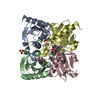
34 subunits of this protein form a single biological assembly. However, this protein adopts two different types of conformations that differ from the deposited crystal structure in the assembly. 23 subunits with one conformation form a ring, and 11 subunits with the other conformation surround the ring in the assembly. Therefore, the biological assembly cannot be described using a simple rotation and translation matrix.

-
要素

#1: タンパク質 Flagellar M-ring protein / FliF


分子量: 19568.713 Da / 分子数: 2 / 断片: UNP residues 58-213 / 由来タイプ: 組換発現
由来: (組換発現) Aquifex aeolicus (strain VF5) (バクテリア)
遺伝子: fliF, aq_1182 / プラスミド: pCold I / 発現宿主: Escherichia coli BL21(DE3) (大腸菌) / 参照: UniProt: O67241
#2: 化合物 ChemComp-MRD / (4R)-2-METHYLPENTANE-2,4-DIOL / (4R)-2-メチル-2,4-ペンタンジオ-ル


分子量: 118.174 Da / 分子数: 1 / 由来タイプ: 合成 / : C6H14O2 / コメント: 沈殿剤*YM
#3: 水 ChemComp-HOH / water


分子量: 18.015 Da / 分子数: 156 / 由来タイプ: 天然 / : H2O
研究の焦点であるリガンドがあるかN
Has protein modificationY

-
実験情報

-
実験

実験手法: X線回折 / 使用した結晶の数: 1

-
試料調製

結晶マシュー密度: 2.61 Å3/Da / 溶媒含有率: 52.94 %
結晶化温度: 277 K / 手法: 蒸気拡散法, ハンギングドロップ法 / pH: 6.2 / 詳細: 0.1M Na-K phosphate pH 6.2, 2.5M NaCl

-
データ収集

回折平均測定温度: 100 K / Serial crystal experiment: N
放射光源由来: シンクロトロン / サイト: SPring-8 / ビームライン: BL41XU / 波長: 1 Å
検出器タイプ: DECTRIS PILATUS3 6M / 検出器: PIXEL / 日付: 2016年10月8日
放射モノクロメーター: Double-crystal monochromator / プロトコル: SINGLE WAVELENGTH / 単色(M)・ラウエ(L): M / 散乱光タイプ: x-ray
放射波長波長: 1 Å / 相対比: 1
反射解像度: 2.29→60.9 Å / Num. obs: 17374 / % possible obs: 96.6 % / 冗長度: 3.2 % / Biso Wilson estimate: 42.18 Å2 / Rmerge(I) obs: 0.054 / Net I/σ(I): 9.8
反射 シェル解像度: 2.29→2.37 Å / Rmerge(I) obs: 0.305 / Mean I/σ(I) obs: 2.9 / Num. unique obs: 1750

-
解析

ソフトウェア
名称バージョン分類
BSSデータ収集
PHENIX1.15.2_3472精密化
MOSFLMデータ削減
Aimlessデータスケーリング
PHENIX位相決定
精密化構造決定の手法: 単波長異常分散 / 解像度: 2.29→60.88 Å / SU ML: 0.358 / 交差検証法: FREE R-VALUE / σ(F): 1.96 / 位相誤差: 28.501
立体化学のターゲット値: GeoStd + Monomer Library + CDL v1.2
Rfactor反射数%反射
Rfree0.2614 879 5.08 %
Rwork0.2132 16441 -
obs0.2155 17320 96.34 %
溶媒の処理減衰半径: 0.9 Å / VDWプローブ半径: 1.11 Å / 溶媒モデル: FLAT BULK SOLVENT MODEL
原子変位パラメータBiso mean: 67.12 Å2
精密化ステップサイクル: LAST / 解像度: 2.29→60.88 Å
タンパク質核酸リガンド溶媒全体
原子数2444 0 8 156 2608
拘束条件
Refine-IDタイプDev ideal
X-RAY DIFFRACTIONf_bond_d0.00382485
X-RAY DIFFRACTIONf_angle_d1.08213350
X-RAY DIFFRACTIONf_chiral_restr0.0431395
X-RAY DIFFRACTIONf_plane_restr0.0042428
X-RAY DIFFRACTIONf_dihedral_angle_d21.2571973
LS精密化 シェル
解像度 (Å)Rfactor RfreeNum. reflection RfreeRfactor RworkNum. reflection RworkRefine-ID% reflection obs (%)
2.29-2.430.351570.28062819X-RAY DIFFRACTION98.54
2.43-2.620.3141730.25762626X-RAY DIFFRACTION94.08
2.62-2.880.29741210.23542798X-RAY DIFFRACTION97.72
2.88-3.30.29491180.23582741X-RAY DIFFRACTION94.61
3.3-4.150.25971720.19192728X-RAY DIFFRACTION97.68
4.15-60.880.21421380.19412729X-RAY DIFFRACTION95.47

+
万見について

-
お知らせ

-
2022年2月9日: EMDBエントリの付随情報ファイルのフォーマットが新しくなりました

EMDBエントリの付随情報ファイルのフォーマットが新しくなりました

  • EMDBのヘッダファイルのバージョン3が、公式のフォーマットとなりました。
  • これまでは公式だったバージョン1.9は、アーカイブから削除されます。

関連情報:EMDBヘッダ

外部リンク:wwPDBはEMDBデータモデルのバージョン3へ移行します

-
2020年8月12日: 新型コロナ情報

新型コロナ情報

URL: https://pdbj.org/emnavi/covid19.php

新ページ: EM Navigatorに新型コロナウイルスの特設ページを開設しました。

関連情報:Covid-19情報 / 2020年3月5日: 新型コロナウイルスの構造データ

+
2020年3月5日: 新型コロナウイルスの構造データ

新型コロナウイルスの構造データ

関連情報:万見生物種 / 2020年8月12日: 新型コロナ情報

外部リンク:COVID-19特集ページ - PDBj / 今月の分子2020年2月:コロナウイルスプロテーアーゼ

+
2019年1月31日: EMDBのIDの桁数の変更

EMDBのIDの桁数の変更

  • EMDBエントリに付与されているアクセスコード(EMDB-ID)は4桁の数字(例、EMD-1234)でしたが、間もなく枯渇します。これまでの4桁のID番号は4桁のまま変更されませんが、4桁の数字を使い切った後に発行されるIDは5桁以上の数字(例、EMD-12345)になります。5桁のIDは2019年の春頃から発行される見通しです。
  • EM Navigator/万見では、接頭語「EMD-」は省略されています。

関連情報:Q: 「EMD」とは何ですか? / 万見/EM NavigatorにおけるID/アクセスコードの表記

外部リンク:EMDB Accession Codes are Changing Soon! / PDBjへお問い合わせ

+
2017年7月12日: PDB大規模アップデート

PDB大規模アップデート

  • 新バージョンのPDBx/mmCIF辞書形式に基づくデータがリリースされました。
  • 今回の更新はバージョン番号が4から5になる大規模なもので、全エントリデータの書き換えが行われる「Remediation」というアップデートに該当します。
  • このバージョンアップで、電子顕微鏡の実験手法に関する多くの項目の書式が改定されました(例:em_softwareなど)。
  • EM NavigatorとYorodumiでも、この改定に基づいた表示内容になります。

外部リンク:wwPDB Remediation / OneDepデータ基準に準拠した、より強化された内容のモデル構造ファイルが、PDBアーカイブで公開されました。

-
万見 (Yorodumi)

幾万の構造データを、幾万の視点から

  • 万見(Yorodumi)は、EMDB/PDB/SASBDBなどの構造データを閲覧するためのページです。
  • EM Navigatorの詳細ページの後継、Omokage検索のフロントエンドも兼ねています。

関連情報:EMDB / PDB / SASBDB / 3つのデータバンクの比較 / 万見検索 / 2016年8月31日: 新しいEM Navigatorと万見 / 万見文献 / Jmol/JSmol / 機能・相同性情報 / 新しいEM Navigatorと万見の変更点

他の情報も見る