 Movie
Movie Controller


+ Open data

- Basic information
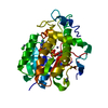
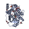
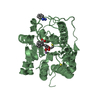
| Entry | Database: PDB / ID: 7cih | ||||||
|---|---|---|---|---|---|---|---|
| Title | Microbial Hormone-sensitive lipase E53 mutant S285G | ||||||
 Components Components | Lipase | ||||||
 Keywords Keywords | HYDROLASE / Esterase / Microbial Hormone-sensitive lipase | ||||||
| Function / homology | : / Alpha/beta hydrolase fold-3 / alpha/beta hydrolase fold / Alpha/Beta hydrolase fold / hydrolase activity / (4-nitrophenyl) hexanoate / 1,4-DIETHYLENE DIOXIDE / P-NITROPHENOL / Lipase Function and homology information Function and homology information | ||||||
| Biological species |  Erythrobacter longus (bacteria) Erythrobacter longus (bacteria) | ||||||
| Method |  X-RAY DIFFRACTION / SYNCHROTRON / MOLECULAR REPLACEMENT / Resolution: 1.789 Å X-RAY DIFFRACTION / SYNCHROTRON / MOLECULAR REPLACEMENT / Resolution: 1.789 Å | ||||||
 Authors Authors | Yang, X. / Li, Z. / Xu, X. / Li, J. | ||||||
| Funding support |  China, 1items China, 1items
| ||||||
 Citation Citation | Journal: Front Microbiol / Year: 2021 Title: Mechanism and Structural Insights Into a Novel Esterase, E53, Isolated From Erythrobacter longus . Authors: Ding, Y. / Nie, L. / Yang, X.C. / Li, Y. / Huo, Y.Y. / Li, Z. / Gao, Y. / Cui, H.L. / Li, J. / Xu, X.W. | ||||||
| History |
|
- Structure visualization
| Structure viewer | Molecule: MolmilJmol/JSmol |
|---|
- Downloads & links
-Download
| PDBx/mmCIF format | 7cih.cif.gz | 278.5 KB | Display | PDBx/mmCIF format |
|---|---|---|---|---|
| PDB format | pdb7cih.ent.gz | 219.3 KB | Display | PDB format |
| PDBx/mmJSON format | 7cih.json.gz | Tree view | PDBx/mmJSON format | |
| Others |  Other downloads Other downloads |
-Validation report
| Arichive directory | https://data.pdbj.org/pub/pdb/validation_reports/ci/7cihftp://data.pdbj.org/pub/pdb/validation_reports/ci/7cih | HTTPS FTP |
|---|
-Related structure data
| Related structure data |  7ci0C  7w8nC 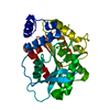 4ypvS S: Starting model for refinement C: citing same article ( |
|---|---|
| Similar structure data |


-Links
 PDBj
PDBj
- Assembly
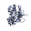
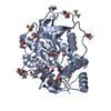
| Deposited unit | 
| |||||||||||||||
|---|---|---|---|---|---|---|---|---|---|---|---|---|---|---|---|---|
| 1 | 
| |||||||||||||||
| 2 | 
| |||||||||||||||
| 3 | 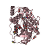
| |||||||||||||||
| 4 | 
| |||||||||||||||
| Unit cell |
| |||||||||||||||
| Components on special symmetry positions |
|
-Components
-Protein , 1 types, 4 molecules ABCD
| #1: Protein | Mass: 33128.703 Da / Num. of mol.: 4 / Mutation: S285G Source method: isolated from a genetically manipulated source Source: (gene. exp.) Erythrobacter longus (bacteria) / Gene: EH31_02760 / Production host: |
|---|
-Non-polymers , 8 types, 1304 molecules 


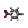


| #2: Chemical | ChemComp-D8F / (  Mass: 237.252 Da / Num. of mol.: 4 / Source method: obtained synthetically / Formula: C12H15NO4 / Feature type: SUBJECT OF INVESTIGATION Mass: 237.252 Da / Num. of mol.: 4 / Source method: obtained synthetically / Formula: C12H15NO4 / Feature type: SUBJECT OF INVESTIGATION#3: Chemical | ChemComp-DMS /  Mass: 78.133 Da / Num. of mol.: 13 / Source method: obtained synthetically / Formula: C2H6OS / Comment: DMSO, precipitant*YM Mass: 78.133 Da / Num. of mol.: 13 / Source method: obtained synthetically / Formula: C2H6OS / Comment: DMSO, precipitant*YM#4: Chemical | ChemComp-GOL /  Mass: 92.094 Da / Num. of mol.: 10 / Source method: obtained synthetically / Formula: C3H8O3 Mass: 92.094 Da / Num. of mol.: 10 / Source method: obtained synthetically / Formula: C3H8O3#5: Chemical | ChemComp-EDO /  Mass: 62.068 Da / Num. of mol.: 18 / Source method: obtained synthetically / Formula: C2H6O2 Mass: 62.068 Da / Num. of mol.: 18 / Source method: obtained synthetically / Formula: C2H6O2#6: Chemical | ChemComp-DIO /  Mass: 88.105 Da / Num. of mol.: 4 / Source method: obtained synthetically / Formula: C4H8O2 Mass: 88.105 Da / Num. of mol.: 4 / Source method: obtained synthetically / Formula: C4H8O2#7: Chemical |  Mass: 139.109 Da / Num. of mol.: 2 / Source method: obtained synthetically / Formula: C6H5NO3 Mass: 139.109 Da / Num. of mol.: 2 / Source method: obtained synthetically / Formula: C6H5NO3#8: Chemical |  Mass: 96.063 Da / Num. of mol.: 2 / Source method: obtained synthetically / Formula: SO4 Mass: 96.063 Da / Num. of mol.: 2 / Source method: obtained synthetically / Formula: SO4#9: Water | ChemComp-HOH / | Mass: 18.015 Da / Num. of mol.: 1251 / Source method: isolated from a natural source / Formula: H2O |
|---|
-Details
| Has ligand of interest | Y |
|---|
-Experimental details
-Experiment
| Experiment | Method: X-RAY DIFFRACTION / Number of used crystals: 1 |
|---|
- Sample preparation
| Crystal | Density Matthews: 3.8 Å3/Da / Density % sol: 67.6 % |
|---|---|
| Crystal grow | Temperature: 291.15 K / Method: vapor diffusion, hanging drop Details: MES pH 6.5, PEG, ammonium sulfate, dioxane, PEG 1000 |
-Data collection
| Diffraction | Mean temperature: 80 K / Serial crystal experiment: N |
|---|---|
| Diffraction source | Source: SYNCHROTRON / Site: SSRF / Beamline: BL19U1 / Wavelength: 0.97776 Å |
| Detector | Type: DECTRIS PILATUS3 S 6M / Detector: PIXEL / Date: May 1, 2017 |
| Radiation | Protocol: SINGLE WAVELENGTH / Monochromatic (M) / Laue (L): M / Scattering type: x-ray |
| Radiation wavelength | Wavelength: 0.97776 Å / Relative weight: 1 |
| Reflection | Resolution: 1.789→48.54 Å / Num. obs: 189466 / % possible obs: 99.47 % / Redundancy: 2 % / Biso Wilson estimate: 30.11 Å2 / CC1/2: 0.999 / CC star: 1 / Rmerge(I) obs: 0.02048 / Net I/σ(I): 18.53 |
| Reflection shell | Resolution: 1.789→1.853 Å / Rmerge(I) obs: 0.3826 / Mean I/σ(I) obs: 2.05 / Num. unique obs: 18072 / CC1/2: 0.825 |
- Processing
| Software |
| ||||||||||||||||
|---|---|---|---|---|---|---|---|---|---|---|---|---|---|---|---|---|---|
| Refinement | Method to determine structure: MOLECULAR REPLACEMENT Starting model: 4YPV Resolution: 1.789→48.54 Å / Cross valid method: FREE R-VALUE Details: Hydrogens have been added in their riding positions
| ||||||||||||||||
| Displacement parameters | Biso mean: 35.88 Å2 | ||||||||||||||||
| Refinement step | Cycle: LAST / Resolution: 1.789→48.54 Å
| ||||||||||||||||
| LS refinement shell | Resolution: 1.789→1.853 Å
|
