 Movie
Movie Controller


+ Open data

- Basic information
| Entry | Database: PDB / ID: 7cen | ||||||
|---|---|---|---|---|---|---|---|
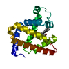
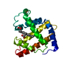
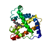
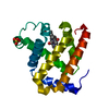
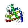
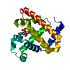
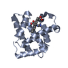
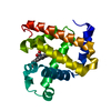
| Title | an x-ray structure of ferric L29E Mb in complex with Mg(II) | ||||||
 Components Components | Myoglobin | ||||||
 Keywords Keywords | OXYGEN STORAGE / myoglobin / sperm whale | ||||||
| Function / homology |  Function and homology information Function and homology informationOxidoreductases; Acting on other nitrogenous compounds as donors / nitrite reductase activity / sarcoplasm / Oxidoreductases; Acting on a peroxide as acceptor; Peroxidases / removal of superoxide radicals / oxygen carrier activity / peroxidase activity / oxygen binding / heme binding / extracellular exosome / metal ion binding Similarity search - Function | ||||||
| Biological species |  | ||||||
| Method |  X-RAY DIFFRACTION / SYNCHROTRON / MOLECULAR REPLACEMENT / Resolution: 2.35 Å X-RAY DIFFRACTION / SYNCHROTRON / MOLECULAR REPLACEMENT / Resolution: 2.35 Å | ||||||
 Authors Authors | Yuan, H. / Lin, Y.W. | ||||||
 Citation Citation | Journal: To Be Published Title: an x-ray structure of ferric L29E Mb in complex with Mg(II) Authors: Yuan, H. / Lin, Y.W. | ||||||
| History |
|
- Structure visualization
| Structure viewer | Molecule: MolmilJmol/JSmol |
|---|
- Downloads & links
-Download
| PDBx/mmCIF format | 7cen.cif.gz | 59.1 KB | Display | PDBx/mmCIF format |
|---|---|---|---|---|
| PDB format | pdb7cen.ent.gz | 33.7 KB | Display | PDB format |
| PDBx/mmJSON format | 7cen.json.gz | Tree view | PDBx/mmJSON format | |
| Others |  Other downloads Other downloads |
-Validation report
| Arichive directory | https://data.pdbj.org/pub/pdb/validation_reports/ce/7cenftp://data.pdbj.org/pub/pdb/validation_reports/ce/7cen | HTTPS FTP |
|---|
-Related structure data
| Related structure data | 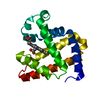 6krcS S: Starting model for refinement |
|---|---|
| Similar structure data |


-Links
 PDBj
PDBj


- Assembly
| Deposited unit | 
| ||||||||||||
|---|---|---|---|---|---|---|---|---|---|---|---|---|---|
| 1 |
| ||||||||||||
| Unit cell |
|
-Components
| #1: Protein | Mass: 17382.104 Da / Num. of mol.: 1 / Mutation: L29E Source method: isolated from a genetically manipulated source Details: SF file contains Friedel pairs. / Source: (gene. exp.)  |
|---|---|
| #2: Chemical | ChemComp-HEM /   Mass: 616.487 Da / Num. of mol.: 1 / Source method: obtained synthetically / Formula: C34H32FeN4O4 Mass: 616.487 Da / Num. of mol.: 1 / Source method: obtained synthetically / Formula: C34H32FeN4O4 |
| #3: Chemical | ChemComp-MG /   Mass: 24.305 Da / Num. of mol.: 1 / Source method: obtained synthetically / Formula: Mg / Feature type: SUBJECT OF INVESTIGATION Mass: 24.305 Da / Num. of mol.: 1 / Source method: obtained synthetically / Formula: Mg / Feature type: SUBJECT OF INVESTIGATION |
| #4: Water | ChemComp-HOH /  Mass: 18.015 Da / Num. of mol.: 112 / Source method: isolated from a natural source / Formula: H2O Mass: 18.015 Da / Num. of mol.: 112 / Source method: isolated from a natural source / Formula: H2O |
| Has ligand of interest | Y |
-Experimental details
-Experiment
| Experiment | Method: X-RAY DIFFRACTION / Number of used crystals: 1 |
|---|
- Sample preparation
| Crystal | Density Matthews: 2.15 Å3/Da / Density % sol: 42.92 % |
|---|---|
| Crystal grow | Temperature: 293 K / Method: vapor diffusion, hanging drop Details: 16% PEG 8000, 0.1 M MES PH 6.0, 0.2 M magnesium chloride |
-Data collection
| Diffraction | Mean temperature: 100 K / Serial crystal experiment: N |
|---|---|
| Diffraction source | Source: SYNCHROTRON / Site: SSRF  / Beamline: BL18U1 / Wavelength: 0.9791 Å / Beamline: BL18U1 / Wavelength: 0.9791 Å |
| Detector | Type: DECTRIS PILATUS 6M / Detector: PIXEL / Date: Jan 10, 2020 |
| Radiation | Protocol: SINGLE WAVELENGTH / Monochromatic (M) / Laue (L): M / Scattering type: x-ray |
| Radiation wavelength | Wavelength: 0.9791 Å / Relative weight: 1 |
| Reflection | Resolution: 2.35→50 Å / Num. obs: 11672 / % possible obs: 99.9 % / Redundancy: 6.7 % / Biso Wilson estimate: 26.87 Å2 / Rmerge(I) obs: 0.164 / Rpim(I) all: 0.068 / Net I/σ(I): 11.3 |
| Reflection shell | Resolution: 2.35→2.39 Å / Rmerge(I) obs: 0.498 / Mean I/σ(I) obs: 4.5 / Num. unique obs: 327 / Rpim(I) all: 0.208 |
- Processing
| Software |
| |||||||||||||||||||||||||||||||||||||||||||||||||||||||||||||||
|---|---|---|---|---|---|---|---|---|---|---|---|---|---|---|---|---|---|---|---|---|---|---|---|---|---|---|---|---|---|---|---|---|---|---|---|---|---|---|---|---|---|---|---|---|---|---|---|---|---|---|---|---|---|---|---|---|---|---|---|---|---|---|---|---|
| Refinement | Method to determine structure: MOLECULAR REPLACEMENT Starting model: 6KRC Resolution: 2.35→38.97 Å / SU ML: 0.2197 / Cross valid method: FREE R-VALUE / σ(F): 1.34 / Phase error: 23.016 Stereochemistry target values: GeoStd + Monomer Library + CDL v1.2
| |||||||||||||||||||||||||||||||||||||||||||||||||||||||||||||||
| Solvent computation | Shrinkage radii: 0.9 Å / VDW probe radii: 1.11 Å / Solvent model: FLAT BULK SOLVENT MODEL | |||||||||||||||||||||||||||||||||||||||||||||||||||||||||||||||
| Displacement parameters | Biso mean: 32.39 Å2 | |||||||||||||||||||||||||||||||||||||||||||||||||||||||||||||||
| Refinement step | Cycle: LAST / Resolution: 2.35→38.97 Å
| |||||||||||||||||||||||||||||||||||||||||||||||||||||||||||||||
| Refine LS restraints |
| |||||||||||||||||||||||||||||||||||||||||||||||||||||||||||||||
| LS refinement shell |
|
