 Movie
Movie Controller

[English] 日本語
 Yorodumi
- PDB-7cc8: Crystal structure of White Spot Syndrome Virus Thymidylate Syntha... -
+ Open data

- Basic information
| Entry | Database: PDB / ID: 7cc8 | ||||||
|---|---|---|---|---|---|---|---|
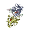
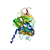
| Title | Crystal structure of White Spot Syndrome Virus Thymidylate Synthase - Apo form | ||||||
 Components Components | Thymidylate Synthase | ||||||
 Keywords Keywords | TRANSFERASE / Thymidylate Synthase / BIOSYNTHETIC PROTEIN | ||||||
| Function / homology |  Function and homology information Function and homology informationthymidylate synthase / thymidylate synthase activity / dTMP biosynthetic process / methylation Similarity search - Function | ||||||
| Biological species |  White spot syndrome virus White spot syndrome virus | ||||||
| Method |  X-RAY DIFFRACTION / SYNCHROTRON / MOLECULAR REPLACEMENT / Resolution: 2.3 Å X-RAY DIFFRACTION / SYNCHROTRON / MOLECULAR REPLACEMENT / Resolution: 2.3 Å | ||||||
 Authors Authors | Kumar, S. / Panchal, N.V. / Shaikh, N. / Vasudevan, D. | ||||||
| Funding support |  India, 1items India, 1items
| ||||||
 Citation Citation | Journal: Int.J.Biol.Macromol. / Year: 2021 Title: Structure analysis of thymidylate synthase from white spot syndrome virus reveals WSSV-specific structural elements. Authors: Panchal, V. / Kumar, S. / Hossain, S.N. / Vasudevan, D. | ||||||
| History |
|
- Structure visualization
| Structure viewer | Molecule: MolmilJmol/JSmol |
|---|
- Downloads & links
-Download
| PDBx/mmCIF format | 7cc8.cif.gz | 130.6 KB | Display | PDBx/mmCIF format |
|---|---|---|---|---|
| PDB format | pdb7cc8.ent.gz | 100.8 KB | Display | PDB format |
| PDBx/mmJSON format | 7cc8.json.gz | Tree view | PDBx/mmJSON format | |
| Others |  Other downloads Other downloads |
-Validation report
| Arichive directory | https://data.pdbj.org/pub/pdb/validation_reports/cc/7cc8ftp://data.pdbj.org/pub/pdb/validation_reports/cc/7cc8 | HTTPS FTP |
|---|
-Related structure data
| Related structure data |  7ccaC  5x5dS S: Starting model for refinement C: citing same article ( |
|---|---|
| Similar structure data |


-Links
 PDBj
PDBj- Assembly
| Deposited unit | 
| ||||||||
|---|---|---|---|---|---|---|---|---|---|
| 1 |
| ||||||||
| Unit cell |
|
-Components
| #1: Protein | Mass: 34193.332 Da / Num. of mol.: 2 Source method: isolated from a genetically manipulated source Source: (gene. exp.) White spot syndrome virus (isolate Shrimp/China/Tongan/1996)Strain: isolate Shrimp/China/Tongan/1996 / Plasmid: pET22b / Production host:  #2: Chemical | ChemComp-SO4 /   Mass: 96.063 Da / Num. of mol.: 6 / Source method: obtained synthetically / Formula: SO4 Mass: 96.063 Da / Num. of mol.: 6 / Source method: obtained synthetically / Formula: SO4#3: Chemical |   Mass: 150.173 Da / Num. of mol.: 3 / Source method: obtained synthetically / Formula: C6H14O4 Mass: 150.173 Da / Num. of mol.: 3 / Source method: obtained synthetically / Formula: C6H14O4#4: Water | ChemComp-HOH / |  Mass: 18.015 Da / Num. of mol.: 164 / Source method: isolated from a natural source / Formula: H2O Mass: 18.015 Da / Num. of mol.: 164 / Source method: isolated from a natural source / Formula: H2OHas ligand of interest | N | |
|---|
-Experimental details
-Experiment
| Experiment | Method: X-RAY DIFFRACTION / Number of used crystals: 1 |
|---|
- Sample preparation
| Crystal | Density Matthews: 3.6 Å3/Da / Density % sol: 65.81 % |
|---|---|
| Crystal grow | Temperature: 291 K / Method: vapor diffusion, sitting drop / pH: 6.5 Details: 200 mM ammonium sulfate, 100 mM Bis-Tris (pH 6.5), 23% PEG3350, 4% Pentaerythritol ethoxylate |
-Data collection
| Diffraction | Mean temperature: 100 K / Serial crystal experiment: N |
|---|---|
| Diffraction source | Source: SYNCHROTRON / Site: ESRF  / Beamline: MASSIF-3 / Wavelength: 0.97242 Å / Beamline: MASSIF-3 / Wavelength: 0.97242 Å |
| Detector | Type: DECTRIS EIGER X 4M / Detector: PIXEL / Date: Sep 1, 2017 |
| Radiation | Protocol: SINGLE WAVELENGTH / Monochromatic (M) / Laue (L): M / Scattering type: x-ray |
| Radiation wavelength | Wavelength: 0.97242 Å / Relative weight: 1 |
| Reflection | Resolution: 2.3→43.41 Å / Num. obs: 42497 / % possible obs: 98.9 % / Redundancy: 5.9 % / Biso Wilson estimate: 38.04 Å2 / CC1/2: 0.999 / Rmerge(I) obs: 0.076 / Net I/σ(I): 15.5 |
| Reflection shell | Resolution: 2.3→2.38 Å / Redundancy: 6.2 % / Rmerge(I) obs: 0.62 / Mean I/σ(I) obs: 2.9 / Num. unique obs: 4356 / CC1/2: 0.807 / % possible all: 99.8 |
- Processing
| Software |
| ||||||||||||||||||
|---|---|---|---|---|---|---|---|---|---|---|---|---|---|---|---|---|---|---|---|
| Refinement | Method to determine structure: MOLECULAR REPLACEMENT Starting model: 5X5D Resolution: 2.3→43.41 Å / Cross valid method: THROUGHOUT
| ||||||||||||||||||
| Displacement parameters | Biso max: 127.32 Å2 / Biso mean: 34.3783 Å2 / Biso min: 19.97 Å2 | ||||||||||||||||||
| Refinement step | Cycle: LAST / Resolution: 2.3→43.41 Å
| ||||||||||||||||||
| LS refinement shell | Resolution: 2.3→2.38 Å
|
