 Movie
Movie Controller

[English] 日本語
 Yorodumi
- PDB-7cbf: Crystal structure of benzophenone synthase from Garcinia mangosta... -
+ Open data

- Basic information
| Entry | Database: PDB / ID: 7cbf | ||||||
|---|---|---|---|---|---|---|---|
| Title | Crystal structure of benzophenone synthase from Garcinia mangostana L. pericarps reveals basis for substrate specificity and catalysis | ||||||
 Components Components | 2,4,6-trihydroxybenzophenone synthase | ||||||
 Keywords Keywords | TRANSFERASE / ACYLTRANSFERASE / BENZOPHENONE SYNTHASE / POLYKETIDE SYNTHASE / GmBPS | ||||||
| Function / homology |  Function and homology information Function and homology information2,4,6-trihydroxybenzophenone synthase / tetrahydroxybenzophenone synthase activity / trihydroxybenzophenone synthase activity / benzoyl-CoA metabolic process / malonyl-CoA metabolic process / polyketide biosynthetic process / acyltransferase activity Similarity search - Function | ||||||
| Biological species |  Garcinia mangostana (mangosteen) Garcinia mangostana (mangosteen) | ||||||
| Method |  X-RAY DIFFRACTION / SYNCHROTRON / MOLECULAR REPLACEMENT / Resolution: 2.301 Å X-RAY DIFFRACTION / SYNCHROTRON / MOLECULAR REPLACEMENT / Resolution: 2.301 Å | ||||||
 Authors Authors | Songsiriritthigul, C. / Nualkaew, N. / Chen, C.-J. | ||||||
 Citation Citation | Journal: Acta Crystallogr.,Sect.F / Year: 2020 Title: Crystal structure of benzophenone synthase from Garcinia mangostana L. pericarps reveals basis for substrate specificity and catalysis. Authors: Songsiriritthigul, C. / Nualkaew, N. / Ketudat-Cairnsb, J. / Chen, C.-J. #1: Journal: Phytochemistry / Year: 2012Title: Benzophenone synthase from Garcinia mangostana L. pericarps Authors: Nualkaew, N. / Morita, H. / Shimokawa, Y. / Kinjo, K. / Kushiro, T. / De-Eknamkul, W. / Ebizuka, Y. / Abe, I. | ||||||
| History |
|
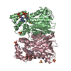
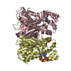
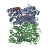
- Structure visualization
| Structure viewer | Molecule: MolmilJmol/JSmol |
|---|
- Downloads & links
-Download
| PDBx/mmCIF format | 7cbf.cif.gz | 169.2 KB | Display | PDBx/mmCIF format |
|---|---|---|---|---|
| PDB format | pdb7cbf.ent.gz | 127.4 KB | Display | PDB format |
| PDBx/mmJSON format | 7cbf.json.gz | Tree view | PDBx/mmJSON format | |
| Others |  Other downloads Other downloads |
-Validation report
| Summary document | 7cbf_validation.pdf.gz | 466.7 KB | Display | wwPDB validaton report |
|---|---|---|---|---|
| Full document | 7cbf_full_validation.pdf.gz | 473.3 KB | Display | |
| Data in XML | 7cbf_validation.xml.gz | 31.6 KB | Display | |
| Data in CIF | 7cbf_validation.cif.gz | 44.7 KB | Display | |
| Arichive directory | https://data.pdbj.org/pub/pdb/validation_reports/cb/7cbfftp://data.pdbj.org/pub/pdb/validation_reports/cb/7cbf | HTTPS FTP |
-Related structure data
| Related structure data |  1cgkS S: Starting model for refinement |
|---|---|
| Similar structure data |


-Links
 PDBj
PDBj


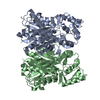
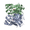
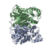
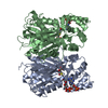
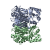
- Assembly
| Deposited unit | 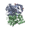
| ||||||||
|---|---|---|---|---|---|---|---|---|---|
| 1 |
| ||||||||
| Unit cell |
|
-Components
| #1: Protein | Mass: 44045.227 Da / Num. of mol.: 2 Source method: isolated from a genetically manipulated source Source: (gene. exp.) Garcinia mangostana (mangosteen) / Gene: BPS / Plasmid: pET22b / Production host:  References: UniProt: L7NCQ3, 2,4,6-trihydroxybenzophenone synthase #2: Chemical |   Mass: 69.085 Da / Num. of mol.: 2 / Source method: obtained synthetically / Formula: C3H5N2 Mass: 69.085 Da / Num. of mol.: 2 / Source method: obtained synthetically / Formula: C3H5N2#3: Chemical | ChemComp-IOD /   Mass: 126.904 Da / Num. of mol.: 10 / Source method: obtained synthetically / Formula: I Mass: 126.904 Da / Num. of mol.: 10 / Source method: obtained synthetically / Formula: I#4: Chemical | ChemComp-GOL /   Mass: 92.094 Da / Num. of mol.: 4 / Source method: obtained synthetically / Formula: C3H8O3 Mass: 92.094 Da / Num. of mol.: 4 / Source method: obtained synthetically / Formula: C3H8O3#5: Water | ChemComp-HOH / |  Mass: 18.015 Da / Num. of mol.: 317 / Source method: isolated from a natural source / Formula: H2O Mass: 18.015 Da / Num. of mol.: 317 / Source method: isolated from a natural source / Formula: H2OHas ligand of interest | N | |
|---|
-Experimental details
-Experiment
| Experiment | Method: X-RAY DIFFRACTION / Number of used crystals: 1 |
|---|
- Sample preparation
| Crystal | Density Matthews: 2.28 Å3/Da / Density % sol: 46.1 % / Description: rectangular morphology |
|---|---|
| Crystal grow | Temperature: 291 K / Method: batch mode / pH: 6.2 / Details: 20% (w/v) PEG 3,350, 0.2 M ammonium iodide, pH 6.2 |
-Data collection
| Diffraction | Mean temperature: 110 K / Serial crystal experiment: N |
|---|---|
| Diffraction source | Source: SYNCHROTRON / Site: NSRRC  / Beamline: BL13C1 / Wavelength: 0.975 Å / Beamline: BL13C1 / Wavelength: 0.975 Å |
| Detector | Type: ADSC QUANTUM 315r / Detector: CCD / Date: Oct 6, 2015 |
| Radiation | Monochromator: DCM / Protocol: SINGLE WAVELENGTH / Monochromatic (M) / Laue (L): M / Scattering type: x-ray |
| Radiation wavelength | Wavelength: 0.975 Å / Relative weight: 1 |
| Reflection | Resolution: 2.3→30 Å / Num. obs: 35272 / % possible obs: 99.5 % / Redundancy: 2.4 % / Biso Wilson estimate: 17.52 Å2 / Rmerge(I) obs: 0.159 / Net I/σ(I): 9.67 |
| Reflection shell | Resolution: 2.3→2.38 Å / Redundancy: 2.4 % / Rmerge(I) obs: 0.49 / Mean I/σ(I) obs: 3.14 / Num. unique obs: 6660 / % possible all: 99.7 |
- Processing
| Software |
| |||||||||||||||||||||||||||||||||||||||||||||||||||||||||||||||||||||||||||||||||||||||||||||||||||||||||||||||||||||||||||||||||||||||||||||||||||||||||||
|---|---|---|---|---|---|---|---|---|---|---|---|---|---|---|---|---|---|---|---|---|---|---|---|---|---|---|---|---|---|---|---|---|---|---|---|---|---|---|---|---|---|---|---|---|---|---|---|---|---|---|---|---|---|---|---|---|---|---|---|---|---|---|---|---|---|---|---|---|---|---|---|---|---|---|---|---|---|---|---|---|---|---|---|---|---|---|---|---|---|---|---|---|---|---|---|---|---|---|---|---|---|---|---|---|---|---|---|---|---|---|---|---|---|---|---|---|---|---|---|---|---|---|---|---|---|---|---|---|---|---|---|---|---|---|---|---|---|---|---|---|---|---|---|---|---|---|---|---|---|---|---|---|---|---|---|---|
| Refinement | Method to determine structure: MOLECULAR REPLACEMENT Starting model: 1CGK Resolution: 2.301→24.347 Å / Cor.coef. Fo:Fc: 0.943 / Cor.coef. Fo:Fc free: 0.91 / SU B: 6.648 / SU ML: 0.161 / Cross valid method: THROUGHOUT / ESU R: 0.339 / ESU R Free: 0.218 Details: Hydrogens have been added in their riding positions
| |||||||||||||||||||||||||||||||||||||||||||||||||||||||||||||||||||||||||||||||||||||||||||||||||||||||||||||||||||||||||||||||||||||||||||||||||||||||||||
| Solvent computation | Ion probe radii: 0.8 Å / Shrinkage radii: 0.8 Å / VDW probe radii: 1.2 Å / Solvent model: MASK BULK SOLVENT | |||||||||||||||||||||||||||||||||||||||||||||||||||||||||||||||||||||||||||||||||||||||||||||||||||||||||||||||||||||||||||||||||||||||||||||||||||||||||||
| Displacement parameters | Biso mean: 16.241 Å2
| |||||||||||||||||||||||||||||||||||||||||||||||||||||||||||||||||||||||||||||||||||||||||||||||||||||||||||||||||||||||||||||||||||||||||||||||||||||||||||
| Refinement step | Cycle: LAST / Resolution: 2.301→24.347 Å
| |||||||||||||||||||||||||||||||||||||||||||||||||||||||||||||||||||||||||||||||||||||||||||||||||||||||||||||||||||||||||||||||||||||||||||||||||||||||||||
| Refine LS restraints |
| |||||||||||||||||||||||||||||||||||||||||||||||||||||||||||||||||||||||||||||||||||||||||||||||||||||||||||||||||||||||||||||||||||||||||||||||||||||||||||
| LS refinement shell |
|
