[English] 日本語
Yorodumi
- PDB-7c8e: Crystal Structure of 14-3-3 epsilon with 9J10 peptide -

+
Open data


ID or keywords:

Loading...

-
Basic information

Entry
Database: PDB / ID: 7c8e
TitleCrystal Structure of 14-3-3 epsilon with 9J10 peptide
Components
  • 14-3-3 protein epsilon
  • 9J10
KeywordsPROTEIN BINDING / Signaling protein / Phosphopeptide binding / peptide complex
Function / homology
Function and homology information


negative regulation of peptidyl-serine dephosphorylation / regulation of heart rate by hormone / regulation of potassium ion transmembrane transporter activity / negative regulation of calcium ion transmembrane transporter activity / membrane repolarization during cardiac muscle cell action potential / negative regulation of toll-like receptor signaling pathway / regulation of membrane repolarization / protein localization to endoplasmic reticulum / NADE modulates death signalling / RAB GEFs exchange GTP for GDP on RABs ...negative regulation of peptidyl-serine dephosphorylation / regulation of heart rate by hormone / regulation of potassium ion transmembrane transporter activity / negative regulation of calcium ion transmembrane transporter activity / membrane repolarization during cardiac muscle cell action potential / negative regulation of toll-like receptor signaling pathway / regulation of membrane repolarization / protein localization to endoplasmic reticulum / NADE modulates death signalling / RAB GEFs exchange GTP for GDP on RABs / Signaling by Hippo / negative regulation of calcium ion export across plasma membrane / cytoplasmic pattern recognition receptor signaling pathway / Deregulated CDK5 triggers multiple neurodegenerative pathways in Alzheimer's disease models / regulation of heart rate by cardiac conduction / protein localization to nucleus / calcium channel regulator activity / phosphoserine residue binding / Regulation of HSF1-mediated heat shock response / HSF1 activation / Activation of BAD and translocation to mitochondria / potassium channel regulator activity / protein targeting / SARS-CoV-2 targets host intracellular signalling and regulatory pathways / Chk1/Chk2(Cds1) mediated inactivation of Cyclin B:Cdk1 complex / signaling adaptor activity / SARS-CoV-1 targets host intracellular signalling and regulatory pathways / RHO GTPases activate PKNs / Loss of Nlp from mitotic centrosomes / Loss of proteins required for interphase microtubule organization from the centrosome / Recruitment of mitotic centrosome proteins and complexes / Recruitment of NuMA to mitotic centrosomes / Anchoring of the basal body to the plasma membrane / protein sequestering activity / regulation of mitotic cell cycle / regulation of cytosolic calcium ion concentration / substantia nigra development / AURKA Activation by TPX2 / positive regulation of protein export from nucleus / Translocation of SLC2A4 (GLUT4) to the plasma membrane / mitochondrial membrane / hippocampus development / TP53 Regulates Metabolic Genes / phosphoprotein binding / neuron migration / cerebral cortex development / histone deacetylase binding / Regulation of PLK1 Activity at G2/M Transition / MAPK cascade / melanosome / MHC class II protein complex binding / cellular response to heat / scaffold protein binding / protein phosphatase binding / transmembrane transporter binding / intracellular signal transduction / cadherin binding / protein heterodimerization activity / protein domain specific binding / focal adhesion / ubiquitin protein ligase binding / enzyme binding / endoplasmic reticulum / signal transduction / RNA binding / extracellular exosome / identical protein binding / membrane / nucleus / cytoplasm / cytosol
Similarity search - Function
14-3-3 proteins signature 2. / 14-3-3 protein, conserved site / 14-3-3 proteins signature 1. / 14-3-3 protein / 14-3-3 homologues / 14-3-3 domain / 14-3-3 domain superfamily / 14-3-3 protein
Similarity search - Domain/homology
14-3-3 protein epsilon
Similarity search - Component
Biological speciesHomo sapiens (human)
Streptomyces avermitilis MA-4680 = NBRC 14893 (bacteria)
MethodX-RAY DIFFRACTION / MOLECULAR REPLACEMENT / Resolution: 3.16 Å
AuthorsMathivanan, S. / Sudhakar, S. / Bairy, S. / Kamariah, N. / Venkitaraman, A.
Funding support India, 1items
OrganizationGrant numberCountry
Department of Biotechnology (DBT, India) India
CitationJournal: Cell Chem Biol / Year: 2021
Title: Target identification for small-molecule discovery in the FOXO3a tumor-suppressor pathway using a biodiverse peptide library.
Authors: Emery, A. / Hardwick, B.S. / Crooks, A.T. / Milech, N. / Watt, P.M. / Mithra, C. / Kumar, V. / Giridharan, S. / Sadasivam, G. / Mathivanan, S. / Sudhakar, S. / Bairy, S. / Bharatham, K. / ...Authors: Emery, A. / Hardwick, B.S. / Crooks, A.T. / Milech, N. / Watt, P.M. / Mithra, C. / Kumar, V. / Giridharan, S. / Sadasivam, G. / Mathivanan, S. / Sudhakar, S. / Bairy, S. / Bharatham, K. / Hurakadli, M.A. / Prasad, T.K. / Kamariah, N. / Muellner, M. / Coelho, M. / Torrance, C.J. / McKenzie, G.J. / Venkitaraman, A.R.
History
DepositionMay 30, 2020Deposition site: PDBJ / Processing site: PDBJ
Revision 1.0Jun 2, 2021Provider: repository / Type: Initial release
Revision 1.1Jun 30, 2021Group: Database references / Category: citation / citation_author
Item: _citation.country / _citation.journal_abbrev ..._citation.country / _citation.journal_abbrev / _citation.journal_id_CSD / _citation.journal_id_ISSN / _citation.pdbx_database_id_DOI / _citation.pdbx_database_id_PubMed / _citation.title / _citation.year
Revision 1.2Dec 8, 2021Group: Database references / Category: citation / database_2
Item: _citation.journal_volume / _citation.page_first ..._citation.journal_volume / _citation.page_first / _citation.page_last / _database_2.pdbx_DOI / _database_2.pdbx_database_accession
Revision 1.3Nov 29, 2023Group: Data collection / Refinement description
Category: chem_comp_atom / chem_comp_bond / pdbx_initial_refinement_model

-
Structure visualization

Structure viewerMolecule:
MolmilJmol/JSmol

Downloads & links

-
Assembly

Deposited unit
A: 14-3-3 protein epsilon
B: 14-3-3 protein epsilon
C: 9J10
D: 9J10
hetero molecules


Theoretical massNumber of molelcules
Total (without water)63,5125
Polymers63,3904
Non-polymers1221
Water52229
1


  • Idetical with deposited unit
  • defined by author&software
  • Evidence: SAXS, HOMO DIMER
TypeNameSymmetry operationNumber
identity operation1_555x,y,z1
Buried area4190 Å2
ΔGint-14 kcal/mol
Surface area22830 Å2
MethodPISA
Unit cell
Length a, b, c (Å)59.470, 83.350, 118.810
Angle α, β, γ (deg.)90.000, 90.000, 90.000
Int Tables number19
Space group name H-MP212121

-
Components

#1: Protein 14-3-3 protein epsilon / 14-3-3E


Mass: 30170.285 Da / Num. of mol.: 2
Source method: isolated from a genetically manipulated source
Source: (gene. exp.) Homo sapiens (human) / Gene: YWHAE / Production host: Escherichia coli BL21(DE3) (bacteria) / Strain (production host): BL21(DE3) / References: UniProt: P62258
#2: Protein/peptide 9J10


Mass: 1524.584 Da / Num. of mol.: 2 / Source method: obtained synthetically
Source: (synth.) Streptomyces avermitilis MA-4680 = NBRC 14893 (bacteria)
#3: Chemical ChemComp-TRS / 2-AMINO-2-HYDROXYMETHYL-PROPANE-1,3-DIOL / TRIS BUFFER


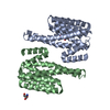
Mass: 122.143 Da / Num. of mol.: 1 / Source method: obtained synthetically / Formula: C4H12NO3 / Feature type: SUBJECT OF INVESTIGATION / Comment: pH buffer*YM
#4: Water ChemComp-HOH / water


Mass: 18.015 Da / Num. of mol.: 29 / Source method: isolated from a natural source / Formula: H2O
Has ligand of interestY

-
Experimental details

-
Experiment

ExperimentMethod: X-RAY DIFFRACTION / Number of used crystals: 1

-
Sample preparation

CrystalDensity Matthews: 2.37 Å3/Da / Density % sol: 48.06 %
Crystal growTemperature: 289.15 K / Method: vapor diffusion, hanging drop / pH: 7.5
Details: 28% PEG 400, 0.1M Hepes pH 7.5, 0.2M Calcium chloride

-
Data collection

DiffractionMean temperature: 100 K / Serial crystal experiment: N
Diffraction sourceSource: ROTATING ANODE / Type: RIGAKU FR-X / Wavelength: 1.5406 Å
DetectorType: RIGAKU RAXIS IV++ / Detector: IMAGE PLATE / Date: Jan 8, 2020
RadiationProtocol: SINGLE WAVELENGTH / Monochromatic (M) / Laue (L): M / Scattering type: x-ray
Radiation wavelengthWavelength: 1.5406 Å / Relative weight: 1
ReflectionResolution: 3.16→68.234 Å / Num. obs: 10688 / % possible obs: 99.9 % / Redundancy: 4.6 % / CC1/2: 0.995 / Rpim(I) all: 0.075 / Rrim(I) all: 0.163 / Rsym value: 0.145 / Net I/σ(I): 8.6
Reflection shellResolution: 3.16→3.33 Å / Redundancy: 4.6 % / Rmerge(I) obs: 0.645 / Mean I/σ(I) obs: 2.3 / Num. unique obs: 1516 / CC1/2: 0.698 / Rpim(I) all: 0.335 / Rrim(I) all: 0.729 / Rsym value: 0.645 / % possible all: 99.5

-
Processing

Software
NameVersionClassification
REFMAC5.8.0257refinement
PDB_EXTRACT3.25data extraction
MOSFLMdata reduction
SCALAdata scaling
PHASERphasing
RefinementMethod to determine structure: MOLECULAR REPLACEMENT
Starting model: 2BR9
Resolution: 3.16→30 Å / Cor.coef. Fo:Fc: 0.926 / Cor.coef. Fo:Fc free: 0.896 / SU B: 26.123 / SU ML: 0.434 / Cross valid method: FREE R-VALUE / σ(F): 0 / ESU R Free: 0.528 / Stereochemistry target values: MAXIMUM LIKELIHOOD
Details: HYDROGENS HAVE BEEN ADDED IN THE RIDING POSITIONS U VALUES : REFINED INDIVIDUALLY
RfactorNum. reflection% reflectionSelection details
Rfree0.2705 522 4.9 %RANDOM
Rwork0.2217 ---
obs0.2242 10115 99.77 %-
Solvent computationIon probe radii: 0.8 Å / Shrinkage radii: 0.8 Å / VDW probe radii: 1.2 Å / Solvent model: MASK
Displacement parametersBiso max: 131.04 Å2 / Biso mean: 62.732 Å2 / Biso min: 23.92 Å2
Baniso -1Baniso -2Baniso -3
1-2.15 Å20 Å20 Å2
2---0.72 Å20 Å2
3----1.43 Å2
Refinement stepCycle: final / Resolution: 3.16→30 Å
ProteinNucleic acidLigandSolventTotal
Num. atoms3692 0 8 29 3729
Biso mean--78.55 39.62 -
Num. residues----473
Refine LS restraints
Refine-IDTypeDev idealDev ideal targetNumber
X-RAY DIFFRACTIONr_bond_refined_d0.0030.0133759
X-RAY DIFFRACTIONr_bond_other_d0.0010.0173463
X-RAY DIFFRACTIONr_angle_refined_deg1.2411.6435088
X-RAY DIFFRACTIONr_angle_other_deg1.1141.587974
X-RAY DIFFRACTIONr_dihedral_angle_1_deg5.9765469
X-RAY DIFFRACTIONr_dihedral_angle_2_deg34.50421.962209
X-RAY DIFFRACTIONr_dihedral_angle_3_deg17.30415648
X-RAY DIFFRACTIONr_dihedral_angle_4_deg13.8891531
X-RAY DIFFRACTIONr_chiral_restr0.0430.2503
X-RAY DIFFRACTIONr_gen_planes_refined0.0030.024245
X-RAY DIFFRACTIONr_gen_planes_other0.0010.02796
LS refinement shellResolution: 3.16→3.236 Å / Rfactor Rfree error: 0
RfactorNum. reflection% reflection
Rfree0.364 33 -
Rwork0.307 724 -
obs--98.83 %

+
About Yorodumi

-
News

-
Feb 9, 2022. New format data for meta-information of EMDB entries

New format data for meta-information of EMDB entries

  • Version 3 of the EMDB header file is now the official format.
  • The previous official version 1.9 will be removed from the archive.

Related info.:EMDB header

External links:wwPDB to switch to version 3 of the EMDB data model

-
Aug 12, 2020. Covid-19 info

Covid-19 info

URL: https://pdbjlvh1.pdbj.org/emnavi/covid19.php

New page: Covid-19 featured information page in EM Navigator.

Related info.:Covid-19 info / Mar 5, 2020. Novel coronavirus structure data

+
Mar 5, 2020. Novel coronavirus structure data

Novel coronavirus structure data

Related info.:Yorodumi Speices / Aug 12, 2020. Covid-19 info

External links:COVID-19 featured content - PDBj / Molecule of the Month (242):Coronavirus Proteases

+
Jan 31, 2019. EMDB accession codes are about to change! (news from PDBe EMDB page)

EMDB accession codes are about to change! (news from PDBe EMDB page)

  • The allocation of 4 digits for EMDB accession codes will soon come to an end. Whilst these codes will remain in use, new EMDB accession codes will include an additional digit and will expand incrementally as the available range of codes is exhausted. The current 4-digit format prefixed with “EMD-” (i.e. EMD-XXXX) will advance to a 5-digit format (i.e. EMD-XXXXX), and so on. It is currently estimated that the 4-digit codes will be depleted around Spring 2019, at which point the 5-digit format will come into force.
  • The EM Navigator/Yorodumi systems omit the EMD- prefix.

Related info.:Q: What is EMD? / ID/Accession-code notation in Yorodumi/EM Navigator

External links:EMDB Accession Codes are Changing Soon! / Contact to PDBj

+
Jul 12, 2017. Major update of PDB

Major update of PDB

  • wwPDB released updated PDB data conforming to the new PDBx/mmCIF dictionary.
  • This is a major update changing the version number from 4 to 5, and with Remediation, in which all the entries are updated.
  • In this update, many items about electron microscopy experimental information are reorganized (e.g. em_software).
  • Now, EM Navigator and Yorodumi are based on the updated data.

External links:wwPDB Remediation / Enriched Model Files Conforming to OneDep Data Standards Now Available in the PDB FTP Archive

-
Yorodumi

Thousand views of thousand structures

  • Yorodumi is a browser for structure data from EMDB, PDB, SASBDB, etc.
  • This page is also the successor to EM Navigator detail page, and also detail information page/front-end page for Omokage search.
  • The word "yorodu" (or yorozu) is an old Japanese word meaning "ten thousand". "mi" (miru) is to see.

Related info.:EMDB / PDB / SASBDB / Comparison of 3 databanks / Yorodumi Search / Aug 31, 2016. New EM Navigator & Yorodumi / Yorodumi Papers / Jmol/JSmol / Function and homology information / Changes in new EM Navigator and Yorodumi

Read more