 Movie
Movie Controller

[English] 日本語
 Yorodumi
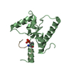
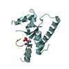
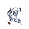
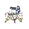
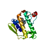
- PDB-7c4q: Crystal structure of DBD plasma treated zebrafish TRF2 myb-domain... -
+ Open data

- Basic information
| Entry | Database: PDB / ID: 7c4q | ||||||
|---|---|---|---|---|---|---|---|
| Title | Crystal structure of DBD plasma treated zebrafish TRF2 myb-domain complexed with DNA | ||||||
 Components Components |
| ||||||
 Keywords Keywords | DNA BINDING PROTEIN / zebrafish TRF2 / telomeric DNA / complex. | ||||||
| Function / homology |  Function and homology information Function and homology informationnegative regulation of telomere maintenance via recombination / telomeric loop formation / cell cycle / negative regulation of telomeric D-loop disassembly / protection from non-homologous end joining at telomere / RNA-templated DNA biosynthetic process / telomeric D-loop disassembly / shelterin complex / regulation of telomere maintenance via telomerase / double-stranded telomeric DNA binding ...negative regulation of telomere maintenance via recombination / telomeric loop formation / cell cycle / negative regulation of telomeric D-loop disassembly / protection from non-homologous end joining at telomere / RNA-templated DNA biosynthetic process / telomeric D-loop disassembly / shelterin complex / regulation of telomere maintenance via telomerase / double-stranded telomeric DNA binding / G-rich strand telomeric DNA binding / protein localization to chromosome, telomeric region / protein homodimerization activity Similarity search - Function | ||||||
| Biological species |   Homo sapiens (human) Homo sapiens (human) | ||||||
| Method |  X-RAY DIFFRACTION / SYNCHROTRON / MOLECULAR REPLACEMENT / Resolution: 2.5 Å X-RAY DIFFRACTION / SYNCHROTRON / MOLECULAR REPLACEMENT / Resolution: 2.5 Å | ||||||
 Authors Authors | Jin, Z. / Park, J.H. / Yun, J.H. / Park, S.Y. / Lee, W. | ||||||
 Citation Citation | Journal: To Be Published Title: Crystal structure of DBD plasma treated zebrafish TRF2 myb-domain complexed with DNA Authors: Jin, Y. / Park, J.H. / Yun, J.H. / Park, S.Y. / Lee, W. | ||||||
| History |
|
- Structure visualization
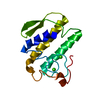
| Structure viewer | Molecule: MolmilJmol/JSmol |
|---|
- Downloads & links
-Download
| PDBx/mmCIF format | 7c4q.cif.gz | 62.2 KB | Display | PDBx/mmCIF format |
|---|---|---|---|---|
| PDB format | pdb7c4q.ent.gz | 40.9 KB | Display | PDB format |
| PDBx/mmJSON format | 7c4q.json.gz | Tree view | PDBx/mmJSON format | |
| Others |  Other downloads Other downloads |
-Validation report
| Summary document | 7c4q_validation.pdf.gz | 419.8 KB | Display | wwPDB validaton report |
|---|---|---|---|---|
| Full document | 7c4q_full_validation.pdf.gz | 419.9 KB | Display | |
| Data in XML | 7c4q_validation.xml.gz | 7.2 KB | Display | |
| Data in CIF | 7c4q_validation.cif.gz | 9.1 KB | Display | |
| Arichive directory | https://data.pdbj.org/pub/pdb/validation_reports/c4/7c4qftp://data.pdbj.org/pub/pdb/validation_reports/c4/7c4q | HTTPS FTP |
-Related structure data
| Related structure data |  7c4pC 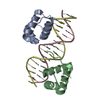 1w0uS S: Starting model for refinement C: citing same article ( |
|---|---|
| Similar structure data |
-Links
 PDBj
PDBj


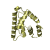
- Assembly
| Deposited unit | 
| ||||||||
|---|---|---|---|---|---|---|---|---|---|
| 1 | 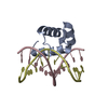
| ||||||||
| 2 | 
| ||||||||
| Unit cell |
|
-Components
-Protein , 2 types, 2 molecules AB
| #1: Protein | Mass: 6521.669 Da / Num. of mol.: 1 Source method: isolated from a genetically manipulated source Source: (gene. exp.)  |
|---|---|
| #4: Protein | Mass: 6620.800 Da / Num. of mol.: 1 Source method: isolated from a genetically manipulated source Source: (gene. exp.) |
-DNA chain , 3 types, 4 molecules CDFE
| #2: DNA chain | Mass: 3773.462 Da / Num. of mol.: 1 / Source method: obtained synthetically / Source: (synth.) Homo sapiens (human) | ||
|---|---|---|---|
| #3: DNA chain | Mass: 3551.346 Da / Num. of mol.: 2 / Source method: obtained synthetically / Source: (synth.) Homo sapiens (human)#5: DNA chain | | Mass: 3115.050 Da / Num. of mol.: 1 / Source method: obtained synthetically / Source: (synth.) Homo sapiens (human) |
-Non-polymers , 1 types, 13 molecules 
| #6: Water | ChemComp-HOH / Mass: 18.015 Da / Num. of mol.: 13 / Source method: isolated from a natural source / Formula: H2O |
|---|
-Experimental details
-Experiment
| Experiment | Method: X-RAY DIFFRACTION / Number of used crystals: 1 |
|---|
- Sample preparation
| Crystal | Density Matthews: 3.15 Å3/Da / Density % sol: 60.98 % |
|---|---|
| Crystal grow | Temperature: 289 K / Method: vapor diffusion, sitting drop / pH: 4.5 Details: 20-25% (w/v) polyethylene glycol 3000, 100 mM Sodium acetate/acetic acid, pH 4.5 |
-Data collection
| Diffraction | Mean temperature: 93 K / Serial crystal experiment: N |
|---|---|
| Diffraction source | Source: SYNCHROTRON / Site: Photon Factory  / Beamline: BL-17A / Wavelength: 1 Å / Beamline: BL-17A / Wavelength: 1 Å |
| Detector | Type: DECTRIS PILATUS3 S 6M / Detector: PIXEL / Date: Nov 25, 2015 |
| Radiation | Protocol: SINGLE WAVELENGTH / Monochromatic (M) / Laue (L): M / Scattering type: x-ray |
| Radiation wavelength | Wavelength: 1 Å / Relative weight: 1 |
| Reflection | Resolution: 2.5→36.297 Å / Num. obs: 12151 / % possible obs: 97.67 % / Redundancy: 7.8 % / Biso Wilson estimate: 57.03 Å2 / Rmerge(I) obs: 0.094 / Net I/σ(I): 16.11 |
| Reflection shell | Resolution: 2.5→2.6 Å / Redundancy: 3.9 % / Rmerge(I) obs: 0.783 / Mean I/σ(I) obs: 0.8 / Num. unique obs: 1097 |
- Processing
| Software |
| ||||||||||||||||||||||||||||||||||||||||||||||||||||||||||||
|---|---|---|---|---|---|---|---|---|---|---|---|---|---|---|---|---|---|---|---|---|---|---|---|---|---|---|---|---|---|---|---|---|---|---|---|---|---|---|---|---|---|---|---|---|---|---|---|---|---|---|---|---|---|---|---|---|---|---|---|---|---|
| Refinement | Method to determine structure: MOLECULAR REPLACEMENT Starting model: 1w0u Resolution: 2.5→36.293 Å / SU ML: 0.42 / Cross valid method: FREE R-VALUE / σ(F): 1.34 / Phase error: 27.97
| ||||||||||||||||||||||||||||||||||||||||||||||||||||||||||||
| Solvent computation | Shrinkage radii: 0.9 Å / VDW probe radii: 1.11 Å | ||||||||||||||||||||||||||||||||||||||||||||||||||||||||||||
| Displacement parameters | Biso max: 89.65 Å2 / Biso mean: 51.7307 Å2 / Biso min: 32.6 Å2 | ||||||||||||||||||||||||||||||||||||||||||||||||||||||||||||
| Refinement step | Cycle: final / Resolution: 2.5→36.293 Å
| ||||||||||||||||||||||||||||||||||||||||||||||||||||||||||||
| Refine LS restraints |
| ||||||||||||||||||||||||||||||||||||||||||||||||||||||||||||
| LS refinement shell | Refine-ID: X-RAY DIFFRACTION / Rfactor Rfree error: 0
|
