 Movie
Movie Controller

[English] 日本語
 Yorodumi
- PDB-7byw: Crystal structure of Acidovorax avenae L-fucose mutarotase (L-fuc... -
+ Open data

- Basic information
| Entry | Database: PDB / ID: 7byw | ||||||
|---|---|---|---|---|---|---|---|
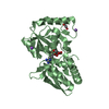
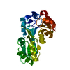
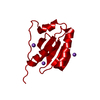
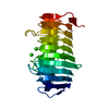
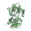
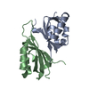
| Title | Crystal structure of Acidovorax avenae L-fucose mutarotase (L-fucose-bound form) | ||||||
 Components Components | L-fucose mutarotase | ||||||
 Keywords Keywords | SUGAR BINDING PROTEIN / L-fucose / Mutarotase | ||||||
| Function / homology | Rhamnose/fucose mutarotase / L-rhamnose mutarotase / racemase and epimerase activity, acting on carbohydrates and derivatives / Dimeric alpha-beta barrel / Chem-1PG / alpha-L-fucopyranose / L-rhamnose mutarotase Function and homology information Function and homology information | ||||||
| Biological species |  Acidovorax avenae subsp. avenae ATCC 19860 (bacteria) Acidovorax avenae subsp. avenae ATCC 19860 (bacteria) | ||||||
| Method |  X-RAY DIFFRACTION / SYNCHROTRON / MOLECULAR REPLACEMENT / Resolution: 1.75 Å X-RAY DIFFRACTION / SYNCHROTRON / MOLECULAR REPLACEMENT / Resolution: 1.75 Å | ||||||
 Authors Authors | Watanabe, Y. / Watanabe, S. | ||||||
 Citation Citation | Journal: Biochem.Biophys.Res.Commun. / Year: 2020 Title: Functional and structural characterization of a novel L-fucose mutarotase involved in non-phosphorylative pathway of L-fucose metabolism. Authors: Watanabe, Y. / Watanabe, S. / Fukui, Y. / Nishiwaki, H. | ||||||
| History |
|
- Structure visualization
| Structure viewer | Molecule: MolmilJmol/JSmol |
|---|
- Downloads & links
-Download
| PDBx/mmCIF format | 7byw.cif.gz | 65.4 KB | Display | PDBx/mmCIF format |
|---|---|---|---|---|
| PDB format | pdb7byw.ent.gz | 46 KB | Display | PDB format |
| PDBx/mmJSON format | 7byw.json.gz | Tree view | PDBx/mmJSON format | |
| Others |  Other downloads Other downloads |
-Validation report
| Arichive directory | https://data.pdbj.org/pub/pdb/validation_reports/by/7bywftp://data.pdbj.org/pub/pdb/validation_reports/by/7byw | HTTPS FTP |
|---|
-Related structure data
| Related structure data | 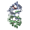 7byuSC S: Starting model for refinement C: citing same article ( |
|---|---|
| Similar structure data |


-Links
 PDBj
PDBj
- Assembly
| Deposited unit | 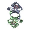
| ||||||||
|---|---|---|---|---|---|---|---|---|---|
| 1 |
| ||||||||
| Unit cell |
|
-Components
| #1: Protein | Mass: 14143.879 Da / Num. of mol.: 2 Source method: isolated from a genetically manipulated source Source: (gene. exp.) Acidovorax avenae subsp. avenae ATCC 19860 (bacteria)Strain: ATCC 19860 / DSM 7227 / JCM 20985 / NCPPB 1011 / Gene: Acav_1655 / Production host: #2: Chemical |   Mass: 252.305 Da / Num. of mol.: 2 / Source method: obtained synthetically / Formula: C11H24O6 Mass: 252.305 Da / Num. of mol.: 2 / Source method: obtained synthetically / Formula: C11H24O6#3: Sugar |   Type: L-saccharide, alpha linking / Mass: 164.156 Da / Num. of mol.: 2 Type: L-saccharide, alpha linking / Mass: 164.156 Da / Num. of mol.: 2Source method: isolated from a genetically manipulated source Formula: C6H12O5 / Feature type: SUBJECT OF INVESTIGATION #4: Water | ChemComp-HOH / |  Mass: 18.015 Da / Num. of mol.: 153 / Source method: isolated from a natural source / Formula: H2O Mass: 18.015 Da / Num. of mol.: 153 / Source method: isolated from a natural source / Formula: H2OHas ligand of interest | Y | |
|---|
-Experimental details
-Experiment
| Experiment | Method: X-RAY DIFFRACTION / Number of used crystals: 1 |
|---|
- Sample preparation
| Crystal | Density Matthews: 3.3 Å3/Da / Density % sol: 62.72 % |
|---|---|
| Crystal grow | Temperature: 293 K / Method: vapor diffusion, sitting drop Details: 0.8 M potassium sodium tartrate, 0.1 M Tris-HCl pH 8.5, 0.5% PEGMME 5000 |
-Data collection
| Diffraction | Mean temperature: 100 K / Serial crystal experiment: N |
|---|---|
| Diffraction source | Source: SYNCHROTRON / Site: SPring-8  / Beamline: BL45XU / Wavelength: 1 Å / Beamline: BL45XU / Wavelength: 1 Å |
| Detector | Type: DECTRIS PILATUS 6M / Detector: PIXEL / Date: Apr 2, 2020 |
| Radiation | Protocol: SINGLE WAVELENGTH / Monochromatic (M) / Laue (L): M / Scattering type: x-ray |
| Radiation wavelength | Wavelength: 1 Å / Relative weight: 1 |
| Reflection | Resolution: 1.75→50 Å / Num. obs: 36466 / % possible obs: 99.9 % / Redundancy: 10.3 % / CC1/2: 0.999 / Rpim(I) all: 0.02 / Rrim(I) all: 0.065 / Rsym value: 0.062 / Net I/σ(I): 17.4 |
| Reflection shell | Resolution: 1.75→1.78 Å / Redundancy: 9.6 % / Mean I/σ(I) obs: 1.9 / Num. unique obs: 1945 / CC1/2: 0.634 / Rpim(I) all: 0.411 / Rrim(I) all: 1.285 / Rsym value: 1.216 / % possible all: 97.8 |
- Processing
| Software |
| ||||||||||||||||||||||||||||||||||||||||||||||||||||||||||||||||||||||||||||||||||||
|---|---|---|---|---|---|---|---|---|---|---|---|---|---|---|---|---|---|---|---|---|---|---|---|---|---|---|---|---|---|---|---|---|---|---|---|---|---|---|---|---|---|---|---|---|---|---|---|---|---|---|---|---|---|---|---|---|---|---|---|---|---|---|---|---|---|---|---|---|---|---|---|---|---|---|---|---|---|---|---|---|---|---|---|---|---|
| Refinement | Method to determine structure: MOLECULAR REPLACEMENT Starting model: 7BYU Resolution: 1.75→44.9876 Å / SU ML: 0.29 / Cross valid method: THROUGHOUT / σ(F): 1.97 / Phase error: 30.51 Details: SF FILE CONTAINS FRIEDEL PAIRS UNDER I/F_MINUS AND I/F_PLUS COLUMNS.
| ||||||||||||||||||||||||||||||||||||||||||||||||||||||||||||||||||||||||||||||||||||
| Solvent computation | Shrinkage radii: 0.9 Å / VDW probe radii: 1.11 Å | ||||||||||||||||||||||||||||||||||||||||||||||||||||||||||||||||||||||||||||||||||||
| Displacement parameters | Biso max: 85.71 Å2 / Biso mean: 41.1657 Å2 / Biso min: 21.02 Å2 | ||||||||||||||||||||||||||||||||||||||||||||||||||||||||||||||||||||||||||||||||||||
| Refinement step | Cycle: final / Resolution: 1.75→44.9876 Å
| ||||||||||||||||||||||||||||||||||||||||||||||||||||||||||||||||||||||||||||||||||||
| LS refinement shell | Refine-ID: X-RAY DIFFRACTION / Rfactor Rfree error: 0
|
