 Movie
Movie Controller

[English] 日本語
 Yorodumi
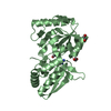
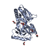
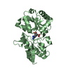
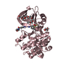
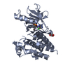
- PDB-5ovz: High resolution structure of the PBP NocT in complex with nopaline -
+ Open data

- Basic information
| Entry | Database: PDB / ID: 5ovz | |||||||||
|---|---|---|---|---|---|---|---|---|---|---|
| Title | High resolution structure of the PBP NocT in complex with nopaline | |||||||||
 Components Components | Nopaline-binding periplasmic protein | |||||||||
 Keywords Keywords | TRANSPORT PROTEIN / periplasmic binding protein | |||||||||
| Function / homology |  Function and homology information Function and homology informationligand-gated monoatomic ion channel activity / outer membrane-bounded periplasmic space / membrane Similarity search - Function | |||||||||
| Biological species |  Agrobacterium (bacteria) Agrobacterium (bacteria) | |||||||||
| Method |  X-RAY DIFFRACTION / SYNCHROTRON / FOURIER SYNTHESIS / Resolution: 1.75 Å X-RAY DIFFRACTION / SYNCHROTRON / FOURIER SYNTHESIS / Resolution: 1.75 Å | |||||||||
 Authors Authors | Vigouroux, A. / Morera, S. | |||||||||
 Citation Citation | Journal: Plos Pathog. / Year: 2014 Title: Agrobacterium uses a unique ligand-binding mode for trapping opines and acquiring a competitive advantage in the niche construction on plant host. Authors: Lang, J. / Vigouroux, A. / Planamente, S. / El Sahili, A. / Blin, P. / Aumont-Nicaise, M. / Dessaux, Y. / Morera, S. / Faure, D. | |||||||||
| History |
|
- Structure visualization
| Structure viewer | Molecule: MolmilJmol/JSmol |
|---|
- Downloads & links
-Download
| PDBx/mmCIF format | 5ovz.cif.gz | 222.2 KB | Display | PDBx/mmCIF format |
|---|---|---|---|---|
| PDB format | pdb5ovz.ent.gz | 176.8 KB | Display | PDB format |
| PDBx/mmJSON format | 5ovz.json.gz | Tree view | PDBx/mmJSON format | |
| Others |  Other downloads Other downloads |
-Validation report
| Arichive directory | https://data.pdbj.org/pub/pdb/validation_reports/ov/5ovzftp://data.pdbj.org/pub/pdb/validation_reports/ov/5ovz | HTTPS FTP |
|---|
-Related structure data
| Related structure data | 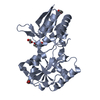 4p0iC 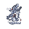 4powC 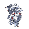 4pp0C  4pox S: Starting model for refinement C: citing same article ( |
|---|---|
| Similar structure data |


-Links
 PDBj
PDBj
- Assembly
| Deposited unit | 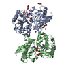
| ||||||||
|---|---|---|---|---|---|---|---|---|---|
| 1 | 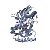
| ||||||||
| 2 | 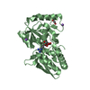
| ||||||||
| Unit cell |
|
-Components
-Protein , 1 types, 2 molecules AB
| #1: Protein | Mass: 29259.627 Da / Num. of mol.: 2 Source method: isolated from a genetically manipulated source Source: (gene. exp.) Agrobacterium (bacteria) / Gene: nocT, Atu6027, AGR_pTi_67 / Production host: |
|---|
-Non-polymers , 5 types, 305 molecules 


| #2: Chemical |  Mass: 304.300 Da / Num. of mol.: 2 / Source method: obtained synthetically / Formula: C11H20N4O6 Mass: 304.300 Da / Num. of mol.: 2 / Source method: obtained synthetically / Formula: C11H20N4O6#3: Chemical |  Mass: 106.120 Da / Num. of mol.: 2 / Source method: obtained synthetically / Formula: C4H10O3 Mass: 106.120 Da / Num. of mol.: 2 / Source method: obtained synthetically / Formula: C4H10O3#4: Chemical | ChemComp-EDO /  Mass: 62.068 Da / Num. of mol.: 6 / Source method: obtained synthetically / Formula: C2H6O2 Mass: 62.068 Da / Num. of mol.: 6 / Source method: obtained synthetically / Formula: C2H6O2#5: Chemical | ChemComp-NA /  Mass: 22.990 Da / Num. of mol.: 7 / Source method: obtained synthetically / Formula: Na Mass: 22.990 Da / Num. of mol.: 7 / Source method: obtained synthetically / Formula: Na#6: Water | ChemComp-HOH / | Mass: 18.015 Da / Num. of mol.: 288 / Source method: isolated from a natural source / Formula: H2O |
|---|
-Experimental details
-Experiment
| Experiment | Method: X-RAY DIFFRACTION / Number of used crystals: 1 |
|---|
- Sample preparation
| Crystal | Density Matthews: 2.44 Å3/Da / Density % sol: 49.62 % |
|---|---|
| Crystal grow | Temperature: 298 K / Method: vapor diffusion, hanging drop / Details: 30% P4000/ 0.1 M Tris pH 8/ 0.1 M LiSO4 |
-Data collection
| Diffraction | Mean temperature: 100 K |
|---|---|
| Diffraction source | Source: SYNCHROTRON / Site: SOLEIL  / Beamline: PROXIMA 1 / Wavelength: 0.97 Å / Beamline: PROXIMA 1 / Wavelength: 0.97 Å |
| Detector | Type: DECTRIS PILATUS3 6M / Detector: PIXEL / Date: Sep 29, 2013 |
| Radiation | Protocol: SINGLE WAVELENGTH / Monochromatic (M) / Laue (L): M / Scattering type: x-ray |
| Radiation wavelength | Wavelength: 0.97 Å / Relative weight: 1 |
| Reflection | Resolution: 1.75→50 Å / Num. all: 55776 / Num. obs: 224742 / % possible obs: 99.8 % / Redundancy: 4 % / Biso Wilson estimate: 33.71 Å2 / CC1/2: 0.999 / Rsym value: 0.05 / Net I/σ(I): 14.18 |
| Reflection shell | Resolution: 1.75→1.86 Å / Mean I/σ(I) obs: 1.41 / Num. measured obs: 35655 / Num. unique all: 8999 / CC1/2: 0.57 / Rsym value: 0.837 / % possible all: 99.5 |
- Processing
| Software |
| ||||||||||||||||||||||||||||||||||||||||||||||||||||||||||||||||||||||||||||||||||||||||||||||||||||||||||||||||||
|---|---|---|---|---|---|---|---|---|---|---|---|---|---|---|---|---|---|---|---|---|---|---|---|---|---|---|---|---|---|---|---|---|---|---|---|---|---|---|---|---|---|---|---|---|---|---|---|---|---|---|---|---|---|---|---|---|---|---|---|---|---|---|---|---|---|---|---|---|---|---|---|---|---|---|---|---|---|---|---|---|---|---|---|---|---|---|---|---|---|---|---|---|---|---|---|---|---|---|---|---|---|---|---|---|---|---|---|---|---|---|---|---|---|---|---|
| Refinement | Method to determine structure: FOURIER SYNTHESIS Starting model: 4POX 4pox Resolution: 1.75→37.4 Å / Cor.coef. Fo:Fc: 0.969 / Cor.coef. Fo:Fc free: 0.961 / Rfactor Rfree error: 0.01 / SU R Cruickshank DPI: 0.101 / Cross valid method: THROUGHOUT / σ(F): 0 / SU R Blow DPI: 0.1 / SU Rfree Blow DPI: 0.091 / SU Rfree Cruickshank DPI: 0.092
| ||||||||||||||||||||||||||||||||||||||||||||||||||||||||||||||||||||||||||||||||||||||||||||||||||||||||||||||||||
| Displacement parameters | Biso mean: 42.15 Å2
| ||||||||||||||||||||||||||||||||||||||||||||||||||||||||||||||||||||||||||||||||||||||||||||||||||||||||||||||||||
| Refine analyze | Luzzati coordinate error obs: 0.2 Å | ||||||||||||||||||||||||||||||||||||||||||||||||||||||||||||||||||||||||||||||||||||||||||||||||||||||||||||||||||
| Refinement step | Cycle: 1 / Resolution: 1.75→37.4 Å
| ||||||||||||||||||||||||||||||||||||||||||||||||||||||||||||||||||||||||||||||||||||||||||||||||||||||||||||||||||
| Refine LS restraints |
| ||||||||||||||||||||||||||||||||||||||||||||||||||||||||||||||||||||||||||||||||||||||||||||||||||||||||||||||||||
| LS refinement shell | Resolution: 1.75→1.79 Å / Rfactor Rfree error: 0 / Total num. of bins used: 20
| ||||||||||||||||||||||||||||||||||||||||||||||||||||||||||||||||||||||||||||||||||||||||||||||||||||||||||||||||||
| Refinement TLS params. | Method: refined / Refine-ID: X-RAY DIFFRACTION
| ||||||||||||||||||||||||||||||||||||||||||||||||||||||||||||||||||||||||||||||||||||||||||||||||||||||||||||||||||
| Refinement TLS group |
|
