 Movie
Movie Controller


+ Open data

- Basic information
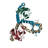
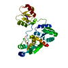
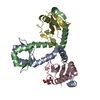
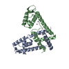
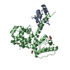
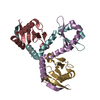
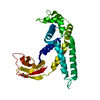
| Entry | Database: PDB / ID: 7bwf | ||||||||||||
|---|---|---|---|---|---|---|---|---|---|---|---|---|---|
| Title | YoeB-YefM complex from Staphylococcus aureus | ||||||||||||
 Components Components |
| ||||||||||||
 Keywords Keywords | TOXIN/ANTITOXIN / Toxin-Antitoxin complex ribosome-dependent ribonuclease ribosome-independent ribonuclease Toxin inhibitor / TOXIN / TOXIN-ANTITOXIN complex | ||||||||||||
| Function / homology |  Function and homology information Function and homology information | ||||||||||||
| Biological species |   Staphylococcus aureus (bacteria) Staphylococcus aureus (bacteria) | ||||||||||||
| Method |  X-RAY DIFFRACTION / SYNCHROTRON / MOLECULAR REPLACEMENT / Resolution: 1.7 Å X-RAY DIFFRACTION / SYNCHROTRON / MOLECULAR REPLACEMENT / Resolution: 1.7 Å | ||||||||||||
 Authors Authors | Lee, B.-J. / Eun, H.-J. | ||||||||||||
| Funding support |  Korea, Republic Of, 3items Korea, Republic Of, 3items
| ||||||||||||
 Citation Citation | Journal: Biochem.Biophys.Res.Commun. / Year: 2020 Title: Crystal structure of the YoeBSa1-YefMSa1 complex from Staphylococcus aureus. Authors: Eun, H.-J. / Lee, K.-Y. / Kim, D.-G. / Im, D.S. / Lee, B.-J. | ||||||||||||
| History |
|
- Structure visualization
| Structure viewer | Molecule: MolmilJmol/JSmol |
|---|
- Downloads & links
-Download
| PDBx/mmCIF format | 7bwf.cif.gz | 223.8 KB | Display | PDBx/mmCIF format |
|---|---|---|---|---|
| PDB format | pdb7bwf.ent.gz | 183.5 KB | Display | PDB format |
| PDBx/mmJSON format | 7bwf.json.gz | Tree view | PDBx/mmJSON format | |
| Others |  Other downloads Other downloads |
-Validation report
| Arichive directory | https://data.pdbj.org/pub/pdb/validation_reports/bw/7bwfftp://data.pdbj.org/pub/pdb/validation_reports/bw/7bwf | HTTPS FTP |
|---|
-Related structure data
| Related structure data |  2a6qS S: Starting model for refinement |
|---|---|
| Similar structure data |

-Links
 PDBj
PDBj- Assembly
| Deposited unit | 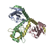
| ||||||||
|---|---|---|---|---|---|---|---|---|---|
| 1 |
| ||||||||
| Unit cell |
|
-Components
| #1: Protein | Mass: 10325.804 Da / Num. of mol.: 2 Source method: isolated from a genetically manipulated source Source: (gene. exp.) Staphylococcus aureus (bacteria) / Gene: M1K003_0948, SAKG03_24000 / Production host: #2: Protein | | Mass: 10335.587 Da / Num. of mol.: 1 Source method: isolated from a genetically manipulated source Source: (gene. exp.) Staphylococcus aureus (bacteria)Gene: yefM, BTN44_14530, EP54_09735, EQ90_00255, HMPREF3211_00052, M1K003_0949, NCTC10654_02579, NCTC10702_03766, NCTC5664_01444, NCTC6133_03236, NCTC7988_02488, RK64_12840, SAKG03_24010 Production host: #3: Protein | | Mass: 10248.509 Da / Num. of mol.: 1 Source method: isolated from a genetically manipulated source Source: (gene. exp.) Staphylococcus aureus (bacteria)Gene: yefM, BTN44_14530, EP54_09735, EQ90_00255, HMPREF3211_00052, M1K003_0949, NCTC10654_02579, NCTC10702_03766, NCTC5664_01444, NCTC6133_03236, NCTC7988_02488, RK64_12840, SAKG03_24010 Production host: #4: Water | ChemComp-HOH / |  Mass: 18.015 Da / Num. of mol.: 174 / Source method: isolated from a natural source / Formula: H2O Mass: 18.015 Da / Num. of mol.: 174 / Source method: isolated from a natural source / Formula: H2O |
|---|
-Experimental details
-Experiment
| Experiment | Method: X-RAY DIFFRACTION / Number of used crystals: 1 |
|---|
- Sample preparation
| Crystal | Density Matthews: 2.24 Å3/Da / Density % sol: 44.97 % |
|---|---|
| Crystal grow | Temperature: 298 K / Method: vapor diffusion, sitting drop Details: 0.08 M sodium chloride, 0.02 M magnesium chloride hexahydrate, 0.04 M sodium cacodylate trihydrate pH 7.0, 40% v/v (+/-)-2-methyl-2,4-pentanediol, and 0.012 M spermine tetrahydrochloride |
-Data collection
| Diffraction | Mean temperature: 100 K / Serial crystal experiment: N |
|---|---|
| Diffraction source | Source: SYNCHROTRON / Site: PAL/PLS / Beamline: 11C / Wavelength: 0.97942 Å |
| Detector | Type: DECTRIS PILATUS3 S 6M / Detector: PIXEL / Date: Jul 31, 2017 |
| Radiation | Protocol: SINGLE WAVELENGTH / Monochromatic (M) / Laue (L): M / Scattering type: x-ray |
| Radiation wavelength | Wavelength: 0.97942 Å / Relative weight: 1 |
| Reflection | Resolution: 1.7→50 Å / Num. obs: 38302 / % possible obs: 97.4 % / Redundancy: 3.4 % / Rmerge(I) obs: 0.069 / Net I/σ(I): 26.9 |
| Reflection shell | Resolution: 1.7→1.73 Å / Rmerge(I) obs: 0.611 / Mean I/σ(I) obs: 2.3 / Num. unique obs: 1944 / % possible all: 99 |
- Processing
| Software |
| ||||||||||||||||||||||||||||||||||||||||||||||||||||||||||||||||||||||||||||||||||||||||||||||||
|---|---|---|---|---|---|---|---|---|---|---|---|---|---|---|---|---|---|---|---|---|---|---|---|---|---|---|---|---|---|---|---|---|---|---|---|---|---|---|---|---|---|---|---|---|---|---|---|---|---|---|---|---|---|---|---|---|---|---|---|---|---|---|---|---|---|---|---|---|---|---|---|---|---|---|---|---|---|---|---|---|---|---|---|---|---|---|---|---|---|---|---|---|---|---|---|---|---|
| Refinement | Method to determine structure: MOLECULAR REPLACEMENT Starting model: 2A6Q Resolution: 1.7→44.832 Å / SU ML: 0.21 / Cross valid method: THROUGHOUT / σ(F): 2 / Phase error: 26.31
| ||||||||||||||||||||||||||||||||||||||||||||||||||||||||||||||||||||||||||||||||||||||||||||||||
| Solvent computation | Shrinkage radii: 0.9 Å / VDW probe radii: 1.11 Å | ||||||||||||||||||||||||||||||||||||||||||||||||||||||||||||||||||||||||||||||||||||||||||||||||
| Displacement parameters | Biso max: 124.07 Å2 / Biso mean: 43.0848 Å2 / Biso min: 18.4 Å2 | ||||||||||||||||||||||||||||||||||||||||||||||||||||||||||||||||||||||||||||||||||||||||||||||||
| Refinement step | Cycle: final / Resolution: 1.7→44.832 Å
| ||||||||||||||||||||||||||||||||||||||||||||||||||||||||||||||||||||||||||||||||||||||||||||||||
| LS refinement shell | Refine-ID: X-RAY DIFFRACTION / Rfactor Rfree error: 0
| ||||||||||||||||||||||||||||||||||||||||||||||||||||||||||||||||||||||||||||||||||||||||||||||||
| Refinement TLS params. | Method: refined / Origin x: 17.3212 Å / Origin y: 9.135 Å / Origin z: 10.9917 Å
| ||||||||||||||||||||||||||||||||||||||||||||||||||||||||||||||||||||||||||||||||||||||||||||||||
| Refinement TLS group |
|
