 Movie
Movie Controller

[English] 日本語
 Yorodumi
- PDB-7btx: The mitochondrial SAM-Mdm10 supercomplex in GDN micelle from S.cere -
+ Open data

- Basic information
| Entry | Database: PDB / ID: 7btx | ||||||||||||||||||||||||||||||||||||
|---|---|---|---|---|---|---|---|---|---|---|---|---|---|---|---|---|---|---|---|---|---|---|---|---|---|---|---|---|---|---|---|---|---|---|---|---|---|
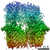
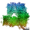
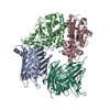
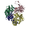
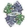
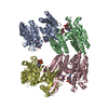
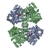
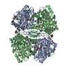
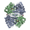
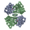
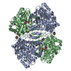
| Title | The mitochondrial SAM-Mdm10 supercomplex in GDN micelle from S.cere | ||||||||||||||||||||||||||||||||||||
 Components Components |
| ||||||||||||||||||||||||||||||||||||
 Keywords Keywords | TRANSLOCASE / Mitochondria | ||||||||||||||||||||||||||||||||||||
| Function / homology |  Function and homology information Function and homology informationERMES complex / mitochondrial outer membrane translocase complex assembly / SAM complex / protein insertion into mitochondrial outer membrane / phospholipid transport / outer membrane / protein import into mitochondrial matrix / mitochondrion organization / mitochondrial outer membrane / mitochondrion Similarity search - Function | ||||||||||||||||||||||||||||||||||||
| Biological species |  | ||||||||||||||||||||||||||||||||||||
| Method | ELECTRON MICROSCOPY / single particle reconstruction / cryo EM / Resolution: 2.8 Å | ||||||||||||||||||||||||||||||||||||
 Authors Authors | Takeda, H. / Tsutsumi, A. / Nishizawa, T. / Nureki, O. / Kikkawa, M. / Endo, T. | ||||||||||||||||||||||||||||||||||||
| Funding support |  Japan, 3items Japan, 3items
| ||||||||||||||||||||||||||||||||||||
 Citation Citation | Journal: Nature / Year: 2021 Title: Mitochondrial sorting and assembly machinery operates by β-barrel switching. Authors: Hironori Takeda / Akihisa Tsutsumi / Tomohiro Nishizawa / Caroline Lindau / Jon V Busto / Lena-Sophie Wenz / Lars Ellenrieder / Kenichiro Imai / Sebastian P Straub / Waltraut Mossmann / Jian ...Authors: Hironori Takeda / Akihisa Tsutsumi / Tomohiro Nishizawa / Caroline Lindau / Jon V Busto / Lena-Sophie Wenz / Lars Ellenrieder / Kenichiro Imai / Sebastian P Straub / Waltraut Mossmann / Jian Qiu / Yu Yamamori / Kentaro Tomii / Junko Suzuki / Takeshi Murata / Satoshi Ogasawara / Osamu Nureki / Thomas Becker / Nikolaus Pfanner / Nils Wiedemann / Masahide Kikkawa / Toshiya Endo /    Abstract: The mitochondrial outer membrane contains so-called β-barrel proteins, which allow communication between the cytosol and the mitochondrial interior. Insertion of β-barrel proteins into the outer ...The mitochondrial outer membrane contains so-called β-barrel proteins, which allow communication between the cytosol and the mitochondrial interior. Insertion of β-barrel proteins into the outer membrane is mediated by the multisubunit mitochondrial sorting and assembly machinery (SAM, also known as TOB). Here we use cryo-electron microscopy to determine the structures of two different forms of the yeast SAM complex at a resolution of 2.8-3.2 Å. The dimeric complex contains two copies of the β-barrel channel protein Sam50-Sam50a and Sam50b-with partially open lateral gates. The peripheral membrane proteins Sam35 and Sam37 cap the Sam50 channels from the cytosolic side, and are crucial for the structural and functional integrity of the dimeric complex. In the second complex, Sam50b is replaced by the β-barrel protein Mdm10. In cooperation with Sam50a, Sam37 recruits and traps Mdm10 by penetrating the interior of its laterally closed β-barrel from the cytosolic side. The substrate-loaded SAM complex contains one each of Sam50, Sam35 and Sam37, but neither Mdm10 nor a second Sam50, suggesting that Mdm10 and Sam50b function as placeholders for a β-barrel substrate released from Sam50a. Our proposed mechanism for dynamic switching of β-barrel subunits and substrate explains how entire precursor proteins can fold in association with the mitochondrial machinery for β-barrel assembly. | ||||||||||||||||||||||||||||||||||||
| History |
|
- Structure visualization
| Movie |
Movie viewer |
|---|---|
| Structure viewer | Molecule: MolmilJmol/JSmol |
- Downloads & links
-Download
| PDBx/mmCIF format | 7btx.cif.gz | 240.5 KB | Display | PDBx/mmCIF format |
|---|---|---|---|---|
| PDB format | pdb7btx.ent.gz | 186.4 KB | Display | PDB format |
| PDBx/mmJSON format | 7btx.json.gz | Tree view | PDBx/mmJSON format | |
| Others |  Other downloads Other downloads |
-Validation report
| Arichive directory | https://data.pdbj.org/pub/pdb/validation_reports/bt/7btxftp://data.pdbj.org/pub/pdb/validation_reports/bt/7btx | HTTPS FTP |
|---|
-Related structure data
| Related structure data |  30190MC 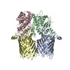 7btwC 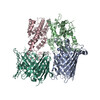 7btyC M: map data used to model this data C: citing same article ( |
|---|---|
| Similar structure data |

-Links
 PDBj
PDBj- Assembly
| Deposited unit | 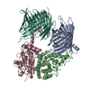
|
|---|---|
| 1 |
|
-Components
| #1: Protein | Mass: 41176.824 Da / Num. of mol.: 1 / Source method: isolated from a natural source / Source: (natural) |
|---|---|
| #2: Protein | Mass: 37446.070 Da / Num. of mol.: 1 / Source method: isolated from a natural source / Source: (natural) |
| #3: Protein | Mass: 37537.797 Da / Num. of mol.: 1 / Source method: isolated from a natural source / Source: (natural) |
| #4: Protein | Mass: 56296.719 Da / Num. of mol.: 1 / Source method: isolated from a natural source / Source: (natural) |
| Has protein modification | N |
-Experimental details
-Experiment
| Experiment | Method: ELECTRON MICROSCOPY |
|---|---|
| EM experiment | Aggregation state: PARTICLE / 3D reconstruction method: single particle reconstruction |
- Sample preparation
| Component | Name: SAM-Mdm10 complex / Type: COMPLEX / Entity ID: all / Source: NATURAL |
|---|---|
| Source (natural) | Organism: |
| Buffer solution | pH: 7.5 |
| Specimen | Embedding applied: NO / Shadowing applied: NO / Staining applied: NO / Vitrification applied: YES |
| Specimen support | Grid material: GOLD / Grid mesh size: 300 divisions/in. / Grid type: C-flat-1.2/1.3 |
| Vitrification | Instrument: FEI VITROBOT MARK IV / Cryogen name: ETHANE / Humidity: 100 % / Chamber temperature: 299 K |
- Electron microscopy imaging
| Experimental equipment |  Model: Titan Krios / Image courtesy: FEI Company |
|---|---|
| Microscopy | Model: FEI TITAN KRIOS |
| Electron gun | Electron source:  FIELD EMISSION GUN / Accelerating voltage: 300 kV / Illumination mode: FLOOD BEAM FIELD EMISSION GUN / Accelerating voltage: 300 kV / Illumination mode: FLOOD BEAM |
| Electron lens | Mode: BRIGHT FIELD |
| Image recording | Electron dose: 60 e/Å2 / Film or detector model: GATAN K3 BIOQUANTUM (6k x 4k) |
- Processing
| Software | Name: PHENIX / Version: 1.14_3260: / Classification: refinement | ||||||||||||||||||||||||||||
|---|---|---|---|---|---|---|---|---|---|---|---|---|---|---|---|---|---|---|---|---|---|---|---|---|---|---|---|---|---|
| EM software |
| ||||||||||||||||||||||||||||
| CTF correction | Type: PHASE FLIPPING AND AMPLITUDE CORRECTION | ||||||||||||||||||||||||||||
| Symmetry | Point symmetry: C1 (asymmetric) | ||||||||||||||||||||||||||||
| 3D reconstruction | Resolution: 2.8 Å / Resolution method: FSC 0.143 CUT-OFF / Num. of particles: 186871 / Symmetry type: POINT |
