Mass: 18.015 Da / Num. of mol.: 966 / Source method: isolated from a natural source / Formula: H2O
-
Experimental details
-
Experiment
Experiment
Method: X-RAY DIFFRACTION / Number of used crystals: 1
-
Sample preparation
Crystal
Density Matthews: 2.76 Å3/Da / Density % sol: 55.5 % / Description: NONE
Crystal grow
Temperature: 293 K / Method: vapor diffusion, hanging drop / pH: 8 Details: CRYSTALS OF THE C. FREUNDII TPL WERE GROWN AT 277 AND 293 K USING THE HANGING DROP VAPOR DIFFUSION METHOD. THE BEST CRYSTALS WERE OBTAINED BY MIXING 2 UL OF THE PROTEIN SOLUTION (18-20 MG/ML) ...Details: CRYSTALS OF THE C. FREUNDII TPL WERE GROWN AT 277 AND 293 K USING THE HANGING DROP VAPOR DIFFUSION METHOD. THE BEST CRYSTALS WERE OBTAINED BY MIXING 2 UL OF THE PROTEIN SOLUTION (18-20 MG/ML) CONTAINING 50MM K-PHOSPHATE PH 8.0, 0.5 MM PLP, 1MM DDT WITH AN EQUAL VOLUME OF THE RESERVOIR SOLUTION CONTAINING 50 MM TRIETHANOLAMINE BUFFER (PH 8.0), 0.5 MM PLP, 2 MM DDT, 0.4 M KCL, AND 35-38% (W/V) POLY(ETHYLENE GLYCOL) 5000 MONOMETHYL ETHER. QUINONOID INTERMEDIATE WITH ALANINE WAS PREPARED BY SOAKING THE TPL CRYSTALS FOR 5 MIN IN THE STABILIZATION SOLUTION CONTAINING 40% POLY(ETHYLENE GLYCOL) 5000 MONOMETHYL ETHER, 50 MM TRIETHANOLAMINE BUFFER (PH 8.0), 0.25 M KCL, 0.2 MM PLP, 0.5 MM DTT AND 10 MM L-ALA.
 Movie
Movie Controller

 Yorodumi
 Open data
 Basic information
 Components
 Keywords
 Function and homology information
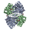
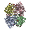
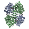
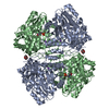
 CITROBACTER FREUNDII (bacteria)
 X-RAY DIFFRACTION /
  Authors
 Citation
 Structure visualization
 Downloads & links
 Other downloads


 PDBj
PDBj Assembly


 Mass: 318.220 Da / Num. of mol.: 2 / Source method: obtained synthetically / Formula: C11H15N2O7P
 Mass: 150.173 Da / Num. of mol.: 1 / Source method: obtained synthetically / Formula: C6H14O4
 Mass: 39.098 Da / Num. of mol.: 2 / Source method: obtained synthetically / Formula: K
 Mass: 326.383 Da / Num. of mol.: 1 / Source method: obtained synthetically / Formula: C14H30O8 / Comment: precipitant*YM
 Sample preparation
 / Beamline: BW7B / Wavelength: 0.862
  Processing