 Movie
Movie Controller


+ Open data

- Basic information
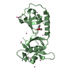
| Entry | Database: PDB / ID: 7bqg | ||||||
|---|---|---|---|---|---|---|---|
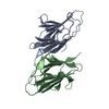
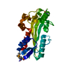
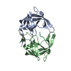
| Title | Complex structure of SAV1 and Dendrin | ||||||
 Components Components | Protein salvador homolog 1,Dendrin | ||||||
 Keywords Keywords | SIGNALING PROTEIN / Hippo pathway / WW domain / SAV1 / Dendrin | ||||||
| Function / homology |  Function and homology information Function and homology informationSignaling by Hippo / intestinal epithelial cell differentiation / regulation of stem cell population maintenance / lung epithelial cell differentiation / positive regulation of hippo signaling / negative regulation of cardiac muscle cell proliferation / hippo signaling / regulation of organ growth / transcription regulator activator activity / dendritic spine membrane ...Signaling by Hippo / intestinal epithelial cell differentiation / regulation of stem cell population maintenance / lung epithelial cell differentiation / positive regulation of hippo signaling / negative regulation of cardiac muscle cell proliferation / hippo signaling / regulation of organ growth / transcription regulator activator activity / dendritic spine membrane / ventricular septum morphogenesis / hair follicle development / keratinocyte differentiation / negative regulation of hippo signaling / positive regulation of fat cell differentiation / cell projection / protein serine/threonine kinase activator activity / negative regulation of epithelial cell proliferation / presynapse / molecular adaptor activity / perikaryon / DNA-binding transcription factor activity, RNA polymerase II-specific / postsynapse / protein stabilization / RNA polymerase II cis-regulatory region sequence-specific DNA binding / positive regulation of apoptotic process / negative regulation of cell population proliferation / apoptotic process / dendrite / endoplasmic reticulum membrane / positive regulation of transcription by RNA polymerase II / identical protein binding / nucleus / plasma membrane / cytosol / cytoplasm Similarity search - Function | ||||||
| Biological species |  | ||||||
| Method |  X-RAY DIFFRACTION / SYNCHROTRON / MOLECULAR REPLACEMENT / Resolution: 1.55010861961 Å X-RAY DIFFRACTION / SYNCHROTRON / MOLECULAR REPLACEMENT / Resolution: 1.55010861961 Å | ||||||
 Authors Authors | Lin, Z. / Zhang, M. | ||||||
| Funding support |  Hong Kong, 1items Hong Kong, 1items
| ||||||
 Citation Citation | Journal: Cell Rep / Year: 2020 Title: A WW Tandem-Mediated Dimerization Mode of SAV1 Essential for Hippo Signaling. Authors: Lin, Z. / Xie, R. / Guan, K. / Zhang, M. | ||||||
| History |
|
- Structure visualization
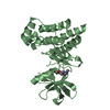
| Structure viewer | Molecule: MolmilJmol/JSmol |
|---|
- Downloads & links
-Download
| PDBx/mmCIF format | 7bqg.cif.gz | 63.8 KB | Display | PDBx/mmCIF format |
|---|---|---|---|---|
| PDB format | pdb7bqg.ent.gz | 37.6 KB | Display | PDB format |
| PDBx/mmJSON format | 7bqg.json.gz | Tree view | PDBx/mmJSON format | |
| Others |  Other downloads Other downloads |
-Validation report
| Arichive directory | https://data.pdbj.org/pub/pdb/validation_reports/bq/7bqgftp://data.pdbj.org/pub/pdb/validation_reports/bq/7bqg | HTTPS FTP |
|---|
-Related structure data
| Related structure data | 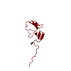 7bqfSC S: Starting model for refinement C: citing same article ( |
|---|---|
| Similar structure data |


-Links
 PDBj
PDBj
- Assembly
| Deposited unit | 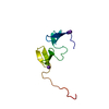
| ||||||||||||
|---|---|---|---|---|---|---|---|---|---|---|---|---|---|
| 1 | 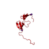
| ||||||||||||
| Unit cell |
| ||||||||||||
| Components on special symmetry positions |
|
-Components
| #1: Protein | Mass: 10320.290 Da / Num. of mol.: 1 Source method: isolated from a genetically manipulated source Source: (gene. exp.)  | ||||
|---|---|---|---|---|---|
| #2: Chemical |   Mass: 39.098 Da / Num. of mol.: 3 / Source method: obtained synthetically / Formula: K / Feature type: SUBJECT OF INVESTIGATION Mass: 39.098 Da / Num. of mol.: 3 / Source method: obtained synthetically / Formula: K / Feature type: SUBJECT OF INVESTIGATION#3: Water | ChemComp-HOH / |  Mass: 18.015 Da / Num. of mol.: 92 / Source method: isolated from a natural source / Formula: H2O Mass: 18.015 Da / Num. of mol.: 92 / Source method: isolated from a natural source / Formula: H2OHas ligand of interest | Y | |
-Experimental details
-Experiment
| Experiment | Method: X-RAY DIFFRACTION / Number of used crystals: 1 |
|---|
- Sample preparation
| Crystal | Density Matthews: 2.01 Å3/Da / Density % sol: 38.88 % |
|---|---|
| Crystal grow | Temperature: 289 K / Method: vapor diffusion, hanging drop Details: 2% w/v PEG 2000 MME and 100 mM BIS-TRIS propane (pH 9.0) |
-Data collection
| Diffraction | Mean temperature: 100 K / Serial crystal experiment: N |
|---|---|
| Diffraction source | Source: SYNCHROTRON / Site: SSRF / Beamline: BL19U1 / Wavelength: 0.97915 Å |
| Detector | Type: DECTRIS PILATUS 6M / Detector: PIXEL / Date: Mar 17, 2019 |
| Radiation | Protocol: SINGLE WAVELENGTH / Monochromatic (M) / Laue (L): M / Scattering type: x-ray |
| Radiation wavelength | Wavelength: 0.97915 Å / Relative weight: 1 |
| Reflection | Resolution: 1.55→50 Å / Num. obs: 11875 / % possible obs: 99.6 % / Redundancy: 12.8 % / Biso Wilson estimate: 12.8744218645 Å2 / CC1/2: 0.99 / Net I/σ(I): 39.1 |
| Reflection shell | Resolution: 1.55→1.58 Å / Num. unique obs: 599 / CC1/2: 0.69 |
- Processing
| Software |
| |||||||||||||||||||||||||||||||||||||||||||||||||||||||||||||||
|---|---|---|---|---|---|---|---|---|---|---|---|---|---|---|---|---|---|---|---|---|---|---|---|---|---|---|---|---|---|---|---|---|---|---|---|---|---|---|---|---|---|---|---|---|---|---|---|---|---|---|---|---|---|---|---|---|---|---|---|---|---|---|---|---|
| Refinement | Method to determine structure: MOLECULAR REPLACEMENT Starting model: 7BQF Resolution: 1.55010861961→49.6925 Å / SU ML: 0.149561517705 / Cross valid method: FREE R-VALUE / σ(F): 0 / Phase error: 21.1462025962
| |||||||||||||||||||||||||||||||||||||||||||||||||||||||||||||||
| Solvent computation | Shrinkage radii: 0.9 Å / VDW probe radii: 1.11 Å | |||||||||||||||||||||||||||||||||||||||||||||||||||||||||||||||
| Displacement parameters | Biso mean: 18.5003240728 Å2 | |||||||||||||||||||||||||||||||||||||||||||||||||||||||||||||||
| Refinement step | Cycle: LAST / Resolution: 1.55010861961→49.6925 Å
| |||||||||||||||||||||||||||||||||||||||||||||||||||||||||||||||
| Refine LS restraints |
| |||||||||||||||||||||||||||||||||||||||||||||||||||||||||||||||
| LS refinement shell |
| |||||||||||||||||||||||||||||||||||||||||||||||||||||||||||||||
| Refinement TLS params. | Method: refined / Origin x: 38.4734067236 Å / Origin y: 12.2274488768 Å / Origin z: 17.0675130407 Å
| |||||||||||||||||||||||||||||||||||||||||||||||||||||||||||||||
| Refinement TLS group | Selection details: all |
