 Movie
Movie Controller

[English] 日本語
 Yorodumi
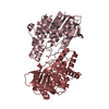
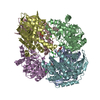
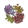
- PDB-7bob: Exo-beta-1,4-mannosidase Op5Man5 from Opitutaceae bacterium strai... -
+ Open data

- Basic information
| Entry | Database: PDB / ID: 7bob | ||||||
|---|---|---|---|---|---|---|---|
| Title | Exo-beta-1,4-mannosidase Op5Man5 from Opitutaceae bacterium strain TAV5 | ||||||
 Components Components | Endo-beta-mannanase | ||||||
 Keywords Keywords | HYDROLASE / Exo-beta-1 / 4-mannosidase / Termite hindgut / Reticulitermes flavipes / Glycosyl hydrolase family 5 / Verrucomicrobia / Opitutaceae | ||||||
| Function / homology |  Function and homology information Function and homology informationClass I glutamine amidotransferase (GATase) domain / Class I glutamine amidotransferase-like / Glycosidases / Glycoside hydrolase superfamily / TIM Barrel / Alpha-Beta Barrel / Rossmann fold / 3-Layer(aba) Sandwich / Alpha Beta Similarity search - Domain/homology | ||||||
| Biological species |  Opitutaceae bacterium TAV5 (bacteria) Opitutaceae bacterium TAV5 (bacteria) | ||||||
| Method |  X-RAY DIFFRACTION / SYNCHROTRON / MOLECULAR REPLACEMENT / Resolution: 2.2 Å X-RAY DIFFRACTION / SYNCHROTRON / MOLECULAR REPLACEMENT / Resolution: 2.2 Å | ||||||
 Authors Authors | Kalyani, D.C. / Reichenbach, T. / Keskitalo, M.M. / Conrad, J. / Aspeborg, H. / Divne, C. | ||||||
| Funding support |  Sweden, 1items Sweden, 1items
| ||||||
 Citation Citation | Journal: J Struct Biol X / Year: 2021 Title: Crystal structure of a homotrimeric verrucomicrobial exo - beta -1,4-mannosidase active in the hindgut of the wood-feeding termite Reticulitermes flavipes . Authors: Kalyani, D.C. / Reichenbach, T. / Keskitalo, M.M. / Conrad, J. / Aspeborg, H. / Divne, C. | ||||||
| History |
|
- Structure visualization
| Structure viewer | Molecule: MolmilJmol/JSmol |
|---|
- Downloads & links
-Download
| PDBx/mmCIF format | 7bob.cif.gz | 332.5 KB | Display | PDBx/mmCIF format |
|---|---|---|---|---|
| PDB format | pdb7bob.ent.gz | 270.2 KB | Display | PDB format |
| PDBx/mmJSON format | 7bob.json.gz | Tree view | PDBx/mmJSON format | |
| Others |  Other downloads Other downloads |
-Validation report
| Summary document | 7bob_validation.pdf.gz | 324.3 KB | Display | wwPDB validaton report |
|---|---|---|---|---|
| Full document | 7bob_full_validation.pdf.gz | 334.1 KB | Display | |
| Data in XML | 7bob_validation.xml.gz | 56.2 KB | Display | |
| Data in CIF | 7bob_validation.cif.gz | 78.7 KB | Display | |
| Arichive directory | https://data.pdbj.org/pub/pdb/validation_reports/bo/7bobftp://data.pdbj.org/pub/pdb/validation_reports/bo/7bob | HTTPS FTP |
-Related structure data
| Similar structure data |
|---|

-Links
 PDBj
PDBj

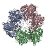
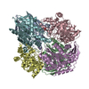
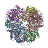
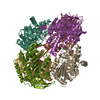
- Assembly
| Deposited unit | 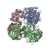
| ||||||||
|---|---|---|---|---|---|---|---|---|---|
| 1 |
| ||||||||
| Unit cell |
|
-Components
| #1: Protein | Mass: 67032.750 Da / Num. of mol.: 3 Source method: isolated from a genetically manipulated source Details: The following regions are flexible and not modeled: residues 213-221; residues 367-380, 588-601 Source: (gene. exp.) Opitutaceae bacterium TAV5 (bacteria) / Gene: OPIT5_10225 / Plasmid: pNIC-CH2 / Production host: #2: Water | ChemComp-HOH / |  Mass: 18.015 Da / Num. of mol.: 189 / Source method: isolated from a natural source / Formula: H2O Mass: 18.015 Da / Num. of mol.: 189 / Source method: isolated from a natural source / Formula: H2O |
|---|
-Experimental details
-Experiment
| Experiment | Method: X-RAY DIFFRACTION / Number of used crystals: 1 |
|---|
- Sample preparation
| Crystal | Density Matthews: 3.47 Å3/Da / Density % sol: 64.54 % |
|---|---|
| Crystal grow | Temperature: 293 K / Method: vapor diffusion, sitting drop / pH: 4.2 Details: 0.1 M phosphorus citrate (pH 4.2), 30 % (v/v) ethanol, and 5 % (w/v) polyethylene glycol 1000 |
-Data collection
| Diffraction | Mean temperature: 100 K / Serial crystal experiment: N |
|---|---|
| Diffraction source | Source: SYNCHROTRON / Site: Diamond  / Beamline: I03 / Wavelength: 0.826561 Å / Beamline: I03 / Wavelength: 0.826561 Å |
| Detector | Type: DECTRIS PILATUS 6M / Detector: PIXEL / Date: Oct 20, 2017 |
| Radiation | Monochromator: Double crystal / Protocol: SINGLE WAVELENGTH / Monochromatic (M) / Laue (L): M / Scattering type: x-ray |
| Radiation wavelength | Wavelength: 0.826561 Å / Relative weight: 1 |
| Reflection | Resolution: 2.2→49.15 Å / Num. obs: 141664 / % possible obs: 99.7 % / Redundancy: 13.3 % / Biso Wilson estimate: 45.16 Å2 / CC1/2: 0.999 / Rsym value: 0.107 / Net I/σ(I): 11.9 |
| Reflection shell | Resolution: 2.2→2.28 Å / Redundancy: 12.6 % / Mean I/σ(I) obs: 1.3 / Num. unique obs: 17459 / CC1/2: 0.768 / Rsym value: 1.865 / % possible all: 99.4 |
- Processing
| Software |
| |||||||||||||||||||||||||||||||||||||||||||||||||||||||||||||||||||||||||||||||||||||||||||||||||||||||||
|---|---|---|---|---|---|---|---|---|---|---|---|---|---|---|---|---|---|---|---|---|---|---|---|---|---|---|---|---|---|---|---|---|---|---|---|---|---|---|---|---|---|---|---|---|---|---|---|---|---|---|---|---|---|---|---|---|---|---|---|---|---|---|---|---|---|---|---|---|---|---|---|---|---|---|---|---|---|---|---|---|---|---|---|---|---|---|---|---|---|---|---|---|---|---|---|---|---|---|---|---|---|---|---|---|---|---|
| Refinement | Method to determine structure: MOLECULAR REPLACEMENT Starting model: Cryo-EM model (3.7 A) Resolution: 2.2→48.867 Å / SU ML: 0.3 / Cross valid method: FREE R-VALUE / σ(F): 1.36 / Phase error: 32.27 / Stereochemistry target values: ML
| |||||||||||||||||||||||||||||||||||||||||||||||||||||||||||||||||||||||||||||||||||||||||||||||||||||||||
| Solvent computation | Shrinkage radii: 0.9 Å / VDW probe radii: 1.11 Å / Solvent model: FLAT BULK SOLVENT MODEL | |||||||||||||||||||||||||||||||||||||||||||||||||||||||||||||||||||||||||||||||||||||||||||||||||||||||||
| Refinement step | Cycle: LAST / Resolution: 2.2→48.867 Å
| |||||||||||||||||||||||||||||||||||||||||||||||||||||||||||||||||||||||||||||||||||||||||||||||||||||||||
| Refine LS restraints |
| |||||||||||||||||||||||||||||||||||||||||||||||||||||||||||||||||||||||||||||||||||||||||||||||||||||||||
| LS refinement shell |
|
