 Movie
Movie Controller

[English] 日本語
 Yorodumi
- PDB-7bnx: Archeal holliday junction resolvase from Thermus thermophilus pha... -
+ Open data

- Basic information
| Entry | Database: PDB / ID: 7bnx | ||||||
|---|---|---|---|---|---|---|---|
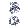
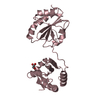
| Title | Archeal holliday junction resolvase from Thermus thermophilus phage 15-6 | ||||||
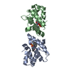
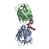
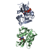
 Components Components | Holliday junction resolvase | ||||||
 Keywords Keywords | RECOMBINATION / archeal holliday junction resolvase helicase DNA binding enzyme phage 15-6 thermus thermophilus | ||||||
| Function / homology | Trna Endonuclease; Chain: A, domain 1 - #10 / Trna Endonuclease; Chain: A, domain 1 / 3-Layer(aba) Sandwich / Alpha Beta Function and homology information Function and homology information | ||||||
| Biological species |   Thermus thermophilus phage 15-6 (virus) Thermus thermophilus phage 15-6 (virus) | ||||||
| Method |  X-RAY DIFFRACTION / SYNCHROTRON / MOLECULAR REPLACEMENT / Resolution: 2.551 Å X-RAY DIFFRACTION / SYNCHROTRON / MOLECULAR REPLACEMENT / Resolution: 2.551 Å | ||||||
 Authors Authors | Hakansson, M. / Ahlqvist, J. / Linares Pasten, J.A. / Jasilionis, A. / Nordberg Karlsson, E. / Al-Karadaghi, S. | ||||||
| Funding support | European Union, 1items
| ||||||
 Citation Citation | Journal: Acta Crystallogr D Struct Biol / Year: 2022 Title: Crystal structure and initial characterization of a novel archaeal-like Holliday junction-resolving enzyme from Thermus thermophilus phage Tth15-6. Authors: Ahlqvist, J. / Linares-Pasten, J.A. / Hakansson, M. / Jasilionis, A. / Kwiatkowska-Semrau, K. / Friðjonsson, O.H. / Kaczorowska, A.K. / Dabrowski, S. / Aevarsson, A. / Hreggvið ...Authors: Ahlqvist, J. / Linares-Pasten, J.A. / Hakansson, M. / Jasilionis, A. / Kwiatkowska-Semrau, K. / Friðjonsson, O.H. / Kaczorowska, A.K. / Dabrowski, S. / Aevarsson, A. / Hreggviðsson, G.O. / Al-Karadaghi, S. / Kaczorowski, T. / Nordberg Karlsson, E. | ||||||
| History |
|
- Structure visualization
| Structure viewer | Molecule: MolmilJmol/JSmol |
|---|
- Downloads & links
-Download
| PDBx/mmCIF format | 7bnx.cif.gz | 117.8 KB | Display | PDBx/mmCIF format |
|---|---|---|---|---|
| PDB format | pdb7bnx.ent.gz | 91.5 KB | Display | PDB format |
| PDBx/mmJSON format | 7bnx.json.gz | Tree view | PDBx/mmJSON format | |
| Others |  Other downloads Other downloads |
-Validation report
| Summary document | 7bnx_validation.pdf.gz | 451.7 KB | Display | wwPDB validaton report |
|---|---|---|---|---|
| Full document | 7bnx_full_validation.pdf.gz | 456.9 KB | Display | |
| Data in XML | 7bnx_validation.xml.gz | 11.7 KB | Display | |
| Data in CIF | 7bnx_validation.cif.gz | 14.9 KB | Display | |
| Arichive directory | https://data.pdbj.org/pub/pdb/validation_reports/bn/7bnxftp://data.pdbj.org/pub/pdb/validation_reports/bn/7bnx | HTTPS FTP |
-Related structure data
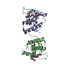
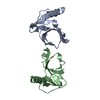
| Related structure data |  7bgsSC S: Starting model for refinement C: citing same article ( |
|---|---|
| Similar structure data |


-Links
 PDBj
PDBj- Assembly
| Deposited unit | 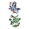
| ||||||||
|---|---|---|---|---|---|---|---|---|---|
| 1 |
| ||||||||
| Unit cell |
| ||||||||
| Components on special symmetry positions |
|
-Components
| #1: Protein | Mass: 18676.576 Da / Num. of mol.: 2 Source method: isolated from a genetically manipulated source Source: (gene. exp.) Thermus thermophilus phage 15-6 (virus)Production host:  #2: Chemical | ChemComp-SO4 /   Mass: 96.063 Da / Num. of mol.: 8 / Source method: obtained synthetically / Formula: SO4 Mass: 96.063 Da / Num. of mol.: 8 / Source method: obtained synthetically / Formula: SO4#3: Water | ChemComp-HOH / |  Mass: 18.015 Da / Num. of mol.: 32 / Source method: isolated from a natural source / Formula: H2O Mass: 18.015 Da / Num. of mol.: 32 / Source method: isolated from a natural source / Formula: H2OHas ligand of interest | N | Has protein modification | Y | |
|---|
-Experimental details
-Experiment
| Experiment | Method: X-RAY DIFFRACTION / Number of used crystals: 1 |
|---|
- Sample preparation
| Crystal | Density Matthews: 2.86 Å3/Da / Density % sol: 57.01 % |
|---|---|
| Crystal grow | Temperature: 293 K / Method: vapor diffusion, sitting drop / pH: 4.2 Details: 1.8 M Ammoniumsulphate and 0.1 M sodiumacetate pH 4.2 |
-Data collection
| Diffraction | Mean temperature: 100 K / Serial crystal experiment: N |
|---|---|
| Diffraction source | Source: SYNCHROTRON / Site: Diamond  / Beamline: I04 / Wavelength: 1.7701 Å / Beamline: I04 / Wavelength: 1.7701 Å |
| Detector | Type: DECTRIS PILATUS3 6M / Detector: PIXEL / Date: Apr 27, 2018 |
| Radiation | Protocol: SINGLE WAVELENGTH / Monochromatic (M) / Laue (L): M / Scattering type: x-ray |
| Radiation wavelength | Wavelength: 1.7701 Å / Relative weight: 1 |
| Reflection | Resolution: 2.55→29 Å / Num. obs: 14269 / % possible obs: 99.8 % / Redundancy: 50.9 % / CC1/2: 1 / Net I/σ(I): 47.2 |
| Reflection shell | Resolution: 2.55→2.66 Å / Rmerge(I) obs: 0.698 / Num. unique obs: 1682 / CC1/2: 0.953 |
- Processing
| Software |
| |||||||||||||||||||||||||||||||||||||||||||||||||||||||||||||||||||||||||||
|---|---|---|---|---|---|---|---|---|---|---|---|---|---|---|---|---|---|---|---|---|---|---|---|---|---|---|---|---|---|---|---|---|---|---|---|---|---|---|---|---|---|---|---|---|---|---|---|---|---|---|---|---|---|---|---|---|---|---|---|---|---|---|---|---|---|---|---|---|---|---|---|---|---|---|---|---|
| Refinement | Method to determine structure: MOLECULAR REPLACEMENT Starting model: 7BGS Resolution: 2.551→28.95 Å / Cor.coef. Fo:Fc: 0.925 / Cor.coef. Fo:Fc free: 0.92 / SU R Cruickshank DPI: 0.334 / Cross valid method: THROUGHOUT / SU R Blow DPI: 0.334 / SU Rfree Blow DPI: 0.241 / SU Rfree Cruickshank DPI: 0.243
| |||||||||||||||||||||||||||||||||||||||||||||||||||||||||||||||||||||||||||
| Displacement parameters | Biso mean: 89.95 Å2
| |||||||||||||||||||||||||||||||||||||||||||||||||||||||||||||||||||||||||||
| Refine analyze | Luzzati coordinate error obs: 0.36 Å | |||||||||||||||||||||||||||||||||||||||||||||||||||||||||||||||||||||||||||
| Refinement step | Cycle: LAST / Resolution: 2.551→28.95 Å
| |||||||||||||||||||||||||||||||||||||||||||||||||||||||||||||||||||||||||||
| Refine LS restraints |
| |||||||||||||||||||||||||||||||||||||||||||||||||||||||||||||||||||||||||||
| LS refinement shell | Resolution: 2.551→2.58 Å
| |||||||||||||||||||||||||||||||||||||||||||||||||||||||||||||||||||||||||||
| Refinement TLS params. | Refine-ID: X-RAY DIFFRACTION
| |||||||||||||||||||||||||||||||||||||||||||||||||||||||||||||||||||||||||||
| Refinement TLS group |
|
