[English] 日本語
Yorodumi
- PDB-7bm8: Crystal structure of the C-terminally truncated chromosome-partit... -

+
Open data


ID or keywords:

Loading...

-
Basic information

Entry
Database: PDB / ID: 7bm8
Title
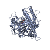
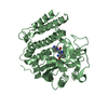
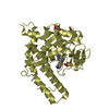
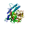
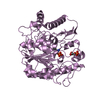
Crystal structure of the C-terminally truncated chromosome-partitioning protein ParB from Caulobacter crescentus complexed with CTP-gamma-S
ComponentsChromosome-partitioning protein ParB
KeywordsDNA BINDING PROTEIN / chromosome segregation / CTP / molecular gates / protein-DNA recognition
Function / homology
Function and homology information


positive regulation of sporulation resulting in formation of a cellular spore / chromosome segregation / chromosome / DNA binding / cytoplasm
Similarity search - Function
ParB/Spo0J, HTH domain / HTH domain found in ParB protein / ParB/RepB/Spo0J partition protein / ParB/Sulfiredoxin domain / ParB/Sulfiredoxin / ParB-like nuclease domain / ParB/Sulfiredoxin superfamily
Similarity search - Domain/homology
CYTIDINE-5'-TRIPHOSPHATE / Chromosome-partitioning protein ParB
Similarity search - Component
Biological speciesCaulobacter vibrioides (bacteria)
MethodX-RAY DIFFRACTION / SYNCHROTRON / MOLECULAR REPLACEMENT / Resolution: 2.73 Å
AuthorsJalal, A.S. / Tran, N.T. / Stevenson, C.E.M. / Lawson, D.M. / Le, T.B.K.
Funding support United Kingdom, 4items
OrganizationGrant numberCountry
Royal SocietyUF140053 United Kingdom
Royal SocietyRG150448 United Kingdom
Biotechnology and Biological Sciences Research Council (BBSRC)BB/P018165/1 United Kingdom
Biotechnology and Biological Sciences Research Council (BBSRC)BBS/E/J/000PR9791 United Kingdom
CitationJournal: Elife / Year: 2021
Title: A CTP-dependent gating mechanism enables ParB spreading on DNA.
Authors: Jalal, A.S. / Tran, N.T. / Stevenson, C.E. / Chimthanawala, A. / Badrinarayanan, A. / Lawson, D.M. / Le, T.B.
History
DepositionJan 19, 2021Deposition site: PDBE / Processing site: PDBE
Revision 1.0Apr 28, 2021Provider: repository / Type: Initial release
Revision 1.1Oct 13, 2021Group: Data collection / Database references
Category: citation / citation_author ...citation / citation_author / database_2 / diffrn_source / pdbx_database_proc
Item: _citation.country / _citation.journal_abbrev ..._citation.country / _citation.journal_abbrev / _citation.journal_id_CSD / _citation.journal_id_ISSN / _citation.journal_volume / _citation.pdbx_database_id_DOI / _citation.pdbx_database_id_PubMed / _citation.title / _citation.year / _database_2.pdbx_DOI / _database_2.pdbx_database_accession / _diffrn_source.pdbx_synchrotron_site
Revision 1.2Jan 31, 2024Group: Data collection / Refinement description
Category: chem_comp_atom / chem_comp_bond ...chem_comp_atom / chem_comp_bond / diffrn_source / pdbx_initial_refinement_model / struct_ncs_dom_lim
Item: _diffrn_source.pdbx_synchrotron_site / _struct_ncs_dom_lim.beg_auth_comp_id ..._diffrn_source.pdbx_synchrotron_site / _struct_ncs_dom_lim.beg_auth_comp_id / _struct_ncs_dom_lim.beg_label_asym_id / _struct_ncs_dom_lim.beg_label_comp_id / _struct_ncs_dom_lim.beg_label_seq_id / _struct_ncs_dom_lim.end_auth_comp_id / _struct_ncs_dom_lim.end_label_asym_id / _struct_ncs_dom_lim.end_label_comp_id / _struct_ncs_dom_lim.end_label_seq_id

-
Structure visualization

Structure viewerMolecule:
MolmilJmol/JSmol

Downloads & links

-
Assembly

Deposited unit
A: Chromosome-partitioning protein ParB
B: Chromosome-partitioning protein ParB
hetero molecules


Theoretical massNumber of molelcules
Total (without water)57,0656
Polymers56,0502
Non-polymers1,0154
Water00
1


  • Idetical with deposited unit
  • defined by author&software
TypeNameSymmetry operationNumber
identity operation1_555x,y,z1
Buried area6340 Å2
ΔGint-50 kcal/mol
Surface area16490 Å2
MethodPISA
Unit cell
Length a, b, c (Å)69.461, 56.076, 71.360
Angle α, β, γ (deg.)90.000, 98.400, 90.000
Int Tables number4
Space group name H-MP1211
Noncrystallographic symmetry (NCS)NCS domain:
IDEns-IDDetails
11A
21B

NCS domain segments:

Component-ID: _ / Ens-ID: 1 / Beg auth comp-ID: GLY / Beg label comp-ID: GLY / End auth comp-ID: GLU / End label comp-ID: GLU / Refine code: _ / Auth seq-ID: 42 - 221 / Label seq-ID: 32 - 211

Dom-IDAuth asym-IDLabel asym-ID
1AA
2BB

-
Components

#1: Protein Chromosome-partitioning protein ParB


Mass: 28024.861 Da / Num. of mol.: 2
Source method: isolated from a genetically manipulated source
Details: The expressed protein comprised residues 11-244 of the wild-type sequence with a C-terminal nickel affinity tag of sequence KLAAALEHHHHHH from the pET21b expression plasmid
Source: (gene. exp.) Caulobacter vibrioides (strain NA1000 / CB15N) (bacteria)
Strain: NA1000 / CB15N / Gene: parB, CCNA_03868 / Production host: Escherichia coli BL21(DE3) (bacteria) / Strain (production host): Rosetta / Variant (production host): Solu / References: UniProt: B8GW30
#2: Chemical ChemComp-CTP / CYTIDINE-5'-TRIPHOSPHATE


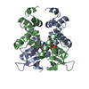
Mass: 483.156 Da / Num. of mol.: 2 / Source method: obtained synthetically / Formula: C9H16N3O14P3 / Feature type: SUBJECT OF INVESTIGATION
#3: Chemical ChemComp-MG / MAGNESIUM ION


Mass: 24.305 Da / Num. of mol.: 2 / Source method: obtained synthetically / Formula: Mg / Feature type: SUBJECT OF INVESTIGATION
Has ligand of interestY

-
Experimental details

-
Experiment

ExperimentMethod: X-RAY DIFFRACTION / Number of used crystals: 1

-
Sample preparation

CrystalDensity Matthews: 2.46 Å3/Da / Density % sol: 50 % / Description: NULL
Crystal growTemperature: 293 K / Method: vapor diffusion, sitting drop / Details: NULL

-
Data collection

DiffractionMean temperature: 100 K / Serial crystal experiment: N
Diffraction sourceSource: SYNCHROTRON / Site: Diamond / Beamline: I03 / Wavelength: 0.976 Å
DetectorType: DECTRIS EIGER2 XE 16M / Detector: PIXEL / Date: Dec 14, 2019
RadiationProtocol: SINGLE WAVELENGTH / Monochromatic (M) / Laue (L): M / Scattering type: x-ray
Radiation wavelengthWavelength: 0.976 Å / Relative weight: 1
ReflectionResolution: 2.73→70.59 Å / Num. obs: 14516 / % possible obs: 98.8 % / Redundancy: 6.4 % / CC1/2: 0.991 / Rmerge(I) obs: 0.195 / Rpim(I) all: 0.083 / Rrim(I) all: 0.212 / Net I/σ(I): 5.4 / Num. measured all: 92266 / Scaling rejects: 77
Reflection shell

Diffraction-ID: 1

Resolution (Å)Redundancy (%)Rmerge(I) obsNum. measured allNum. unique obsCC1/2Rpim(I) allRrim(I) allNet I/σ(I) obs% possible all
2.73-2.864.81.21847317560.8250.5991.3571.291.4
9.05-70.595.90.06325614320.9910.0280.06913.599.7

-
Processing

Software
NameVersionClassification
Aimless0.7.4data scaling
REFMAC5.8.0258refinement
PDB_EXTRACT3.25data extraction
DIALSdata reduction
PHASERphasing
RefinementMethod to determine structure: MOLECULAR REPLACEMENT
Starting model: 6T1F
Resolution: 2.73→68.81 Å / Cor.coef. Fo:Fc: 0.93 / Cor.coef. Fo:Fc free: 0.911 / SU B: 21.471 / SU ML: 0.384 / SU R Cruickshank DPI: 0.6609 / Cross valid method: THROUGHOUT / σ(F): 0 / ESU R: 0.661 / ESU R Free: 0.351 / Stereochemistry target values: MAXIMUM LIKELIHOOD
Details: HYDROGENS HAVE BEEN ADDED IN THE RIDING POSITIONS. The density did not allow the unambiguous placement of the gamma-S of CTP-gamma-S. Thus CTP was refined in its place.
RfactorNum. reflection% reflectionSelection details
Rfree0.2839 678 4.7 %RANDOM
Rwork0.2477 ---
obs0.2493 13824 98.65 %-
Solvent computationIon probe radii: 0.7 Å / Shrinkage radii: 0.7 Å / VDW probe radii: 1.1 Å / Solvent model: MASK
Displacement parametersBiso max: 186.77 Å2 / Biso mean: 68.899 Å2 / Biso min: 33.59 Å2
Baniso -1Baniso -2Baniso -3
1--6.79 Å20 Å21.2 Å2
2---6.21 Å20 Å2
3---12.12 Å2
Refinement stepCycle: final / Resolution: 2.73→68.81 Å
ProteinNucleic acidLigandSolventTotal
Num. atoms2656 0 60 0 2716
Biso mean--60.63 --
Num. residues----360
Refine LS restraints
Refine-IDTypeDev idealDev ideal targetNumber
X-RAY DIFFRACTIONr_bond_refined_d0.0020.0132756
X-RAY DIFFRACTIONr_bond_other_d0.0010.0172521
X-RAY DIFFRACTIONr_angle_refined_deg1.1881.6513743
X-RAY DIFFRACTIONr_angle_other_deg1.1051.5775807
X-RAY DIFFRACTIONr_dihedral_angle_1_deg4.8895358
X-RAY DIFFRACTIONr_dihedral_angle_2_deg25.97321.2150
X-RAY DIFFRACTIONr_dihedral_angle_3_deg15.52815437
X-RAY DIFFRACTIONr_dihedral_angle_4_deg11.4031530
X-RAY DIFFRACTIONr_chiral_restr0.0330.2386
X-RAY DIFFRACTIONr_gen_planes_refined0.0020.023126
X-RAY DIFFRACTIONr_gen_planes_other0.0010.02546
Refine LS restraints NCS

Ens-ID: 1 / Number: 5284 / Refine-ID: X-RAY DIFFRACTION / Type: interatomic distance / Rms dev position: 0.05 Å / Weight position: 0.05

Dom-IDAuth asym-ID
1A
2B
LS refinement shellResolution: 2.73→2.8 Å / Rfactor Rfree error: 0
RfactorNum. reflection% reflection
Rfree0.405 61 -
Rwork0.371 870 -
obs--84.95 %

+
About Yorodumi

-
News

-
Feb 9, 2022. New format data for meta-information of EMDB entries

New format data for meta-information of EMDB entries

  • Version 3 of the EMDB header file is now the official format.
  • The previous official version 1.9 will be removed from the archive.

Related info.:EMDB header

External links:wwPDB to switch to version 3 of the EMDB data model

-
Aug 12, 2020. Covid-19 info

Covid-19 info

URL: https://pdbjlvh1.pdbj.org/emnavi/covid19.php

New page: Covid-19 featured information page in EM Navigator.

Related info.:Covid-19 info / Mar 5, 2020. Novel coronavirus structure data

+
Mar 5, 2020. Novel coronavirus structure data

Novel coronavirus structure data

Related info.:Yorodumi Speices / Aug 12, 2020. Covid-19 info

External links:COVID-19 featured content - PDBj / Molecule of the Month (242):Coronavirus Proteases

+
Jan 31, 2019. EMDB accession codes are about to change! (news from PDBe EMDB page)

EMDB accession codes are about to change! (news from PDBe EMDB page)

  • The allocation of 4 digits for EMDB accession codes will soon come to an end. Whilst these codes will remain in use, new EMDB accession codes will include an additional digit and will expand incrementally as the available range of codes is exhausted. The current 4-digit format prefixed with “EMD-” (i.e. EMD-XXXX) will advance to a 5-digit format (i.e. EMD-XXXXX), and so on. It is currently estimated that the 4-digit codes will be depleted around Spring 2019, at which point the 5-digit format will come into force.
  • The EM Navigator/Yorodumi systems omit the EMD- prefix.

Related info.:Q: What is EMD? / ID/Accession-code notation in Yorodumi/EM Navigator

External links:EMDB Accession Codes are Changing Soon! / Contact to PDBj

+
Jul 12, 2017. Major update of PDB

Major update of PDB

  • wwPDB released updated PDB data conforming to the new PDBx/mmCIF dictionary.
  • This is a major update changing the version number from 4 to 5, and with Remediation, in which all the entries are updated.
  • In this update, many items about electron microscopy experimental information are reorganized (e.g. em_software).
  • Now, EM Navigator and Yorodumi are based on the updated data.

External links:wwPDB Remediation / Enriched Model Files Conforming to OneDep Data Standards Now Available in the PDB FTP Archive

-
Yorodumi

Thousand views of thousand structures

  • Yorodumi is a browser for structure data from EMDB, PDB, SASBDB, etc.
  • This page is also the successor to EM Navigator detail page, and also detail information page/front-end page for Omokage search.
  • The word "yorodu" (or yorozu) is an old Japanese word meaning "ten thousand". "mi" (miru) is to see.

Related info.:EMDB / PDB / SASBDB / Comparison of 3 databanks / Yorodumi Search / Aug 31, 2016. New EM Navigator & Yorodumi / Yorodumi Papers / Jmol/JSmol / Function and homology information / Changes in new EM Navigator and Yorodumi

Read more