 Movie
Movie Controller

[English] 日本語
 Yorodumi
- PDB-7bhm: Crystal structure of hen egg white lysozyme using drop-on-drop SF... -
+ Open data

- Basic information
| Entry | Database: PDB / ID: 7bhm | ||||||||||||
|---|---|---|---|---|---|---|---|---|---|---|---|---|---|
| Title | Crystal structure of hen egg white lysozyme using drop-on-drop SFX method - 0.7 s mixing with N-acetyl-D-glucosamine. | ||||||||||||
 Components Components | Lysozyme | ||||||||||||
 Keywords Keywords | HYDROLASE / lysozyme / SFX / XFEL | ||||||||||||
| Function / homology |  Function and homology information Function and homology informationLactose synthesis / Antimicrobial peptides / Neutrophil degranulation / beta-N-acetylglucosaminidase activity / cell wall macromolecule catabolic process / lysozyme / lysozyme activity / killing of cells of another organism / defense response to Gram-negative bacterium / defense response to bacterium ...Lactose synthesis / Antimicrobial peptides / Neutrophil degranulation / beta-N-acetylglucosaminidase activity / cell wall macromolecule catabolic process / lysozyme / lysozyme activity / killing of cells of another organism / defense response to Gram-negative bacterium / defense response to bacterium / defense response to Gram-positive bacterium / endoplasmic reticulum / : / identical protein binding / cytoplasm Similarity search - Function | ||||||||||||
| Biological species |  | ||||||||||||
| Method |  X-RAY DIFFRACTION / FREE ELECTRON LASER / MOLECULAR REPLACEMENT / Resolution: 1.45 Å X-RAY DIFFRACTION / FREE ELECTRON LASER / MOLECULAR REPLACEMENT / Resolution: 1.45 Å | ||||||||||||
 Authors Authors | Butryn, A. / Orville, A.M. | ||||||||||||
| Funding support |  United Kingdom, 3items United Kingdom, 3items
| ||||||||||||
 Citation Citation | Journal: Nat Commun / Year: 2021 Title: An on-demand, drop-on-drop method for studying enzyme catalysis by serial crystallography. Authors: Butryn, A. / Simon, P.S. / Aller, P. / Hinchliffe, P. / Massad, R.N. / Leen, G. / Tooke, C.L. / Bogacz, I. / Kim, I.S. / Bhowmick, A. / Brewster, A.S. / Devenish, N.E. / Brem, J. / Kamps, J. ...Authors: Butryn, A. / Simon, P.S. / Aller, P. / Hinchliffe, P. / Massad, R.N. / Leen, G. / Tooke, C.L. / Bogacz, I. / Kim, I.S. / Bhowmick, A. / Brewster, A.S. / Devenish, N.E. / Brem, J. / Kamps, J.J.A.G. / Lang, P.A. / Rabe, P. / Axford, D. / Beale, J.H. / Davy, B. / Ebrahim, A. / Orlans, J. / Storm, S.L.S. / Zhou, T. / Owada, S. / Tanaka, R. / Tono, K. / Evans, G. / Owen, R.L. / Houle, F.A. / Sauter, N.K. / Schofield, C.J. / Spencer, J. / Yachandra, V.K. / Yano, J. / Kern, J.F. / Orville, A.M. | ||||||||||||
| History |
|
- Structure visualization
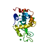
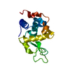
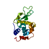
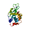
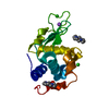
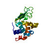
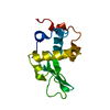
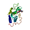
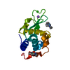
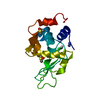
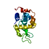
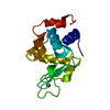
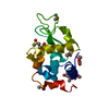
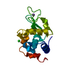
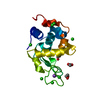
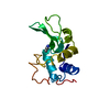
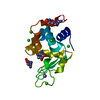
| Structure viewer | Molecule: MolmilJmol/JSmol |
|---|
- Downloads & links
-Download
| PDBx/mmCIF format | 7bhm.cif.gz | 51.7 KB | Display | PDBx/mmCIF format |
|---|---|---|---|---|
| PDB format | pdb7bhm.ent.gz | 28.6 KB | Display | PDB format |
| PDBx/mmJSON format | 7bhm.json.gz | Tree view | PDBx/mmJSON format | |
| Others |  Other downloads Other downloads |
-Validation report
| Arichive directory | https://data.pdbj.org/pub/pdb/validation_reports/bh/7bhmftp://data.pdbj.org/pub/pdb/validation_reports/bh/7bhm | HTTPS FTP |
|---|
-Related structure data
| Related structure data |  7bh3C  7bh4C 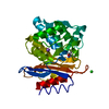 7bh5C 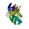 7bh6C 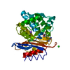 7bh7C  7bhkC  7bhlC  7bhnC 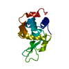 4etaS C: citing same article ( S: Starting model for refinement |
|---|---|
| Similar structure data |


-Links
 PDBj
PDBj


- Assembly
| Deposited unit | 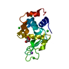
| ||||||||||||
|---|---|---|---|---|---|---|---|---|---|---|---|---|---|
| 1 |
| ||||||||||||
| Unit cell |
| ||||||||||||
| Components on special symmetry positions |
|
-Components
| #1: Protein | Mass: 14331.160 Da / Num. of mol.: 1 / Source method: isolated from a natural source / Source: (natural) | ||||||||
|---|---|---|---|---|---|---|---|---|---|
| #2: Sugar | ChemComp-NDG /   Type: D-saccharide, alpha linking / Mass: 221.208 Da / Num. of mol.: 1 / Source method: obtained synthetically / Formula: C8H15NO6 / Feature type: SUBJECT OF INVESTIGATION Type: D-saccharide, alpha linking / Mass: 221.208 Da / Num. of mol.: 1 / Source method: obtained synthetically / Formula: C8H15NO6 / Feature type: SUBJECT OF INVESTIGATION | ||||||||
| #3: Chemical | ChemComp-CL /   Mass: 35.453 Da / Num. of mol.: 4 / Source method: obtained synthetically / Formula: Cl Mass: 35.453 Da / Num. of mol.: 4 / Source method: obtained synthetically / Formula: Cl#4: Chemical | ChemComp-NA / |   Mass: 22.990 Da / Num. of mol.: 1 / Source method: obtained synthetically / Formula: Na Mass: 22.990 Da / Num. of mol.: 1 / Source method: obtained synthetically / Formula: Na#5: Water | ChemComp-HOH / |  Mass: 18.015 Da / Num. of mol.: 83 / Source method: isolated from a natural source / Formula: H2O Mass: 18.015 Da / Num. of mol.: 83 / Source method: isolated from a natural source / Formula: H2OHas ligand of interest | Y | Has protein modification | Y | |
-Experimental details
-Experiment
| Experiment | Method: X-RAY DIFFRACTION / Number of used crystals: 1 |
|---|
- Sample preparation
| Crystal | Density Matthews: 2.06 Å3/Da / Density % sol: 40.25 % |
|---|---|
| Crystal grow | Temperature: 295 K / Method: batch mode Details: 1 M citric acid, 20% (w/v) NaCl, 5% (w/v) PEG 6000, pH 3.0 |
-Data collection
| Diffraction | Mean temperature: 308 K / Serial crystal experiment: Y |
|---|---|
| Diffraction source | Source: FREE ELECTRON LASER / Site: SACLA  / Beamline: BL2 / Wavelength: 1.24 Å / Beamline: BL2 / Wavelength: 1.24 Å |
| Detector | Type: RAYONIX MX300-HS / Detector: CCD / Date: Jul 11, 2019 |
| Radiation | Protocol: SINGLE WAVELENGTH / Monochromatic (M) / Laue (L): M / Scattering type: x-ray |
| Radiation wavelength | Wavelength: 1.24 Å / Relative weight: 1 |
| Reflection | Resolution: 1.45→55.72 Å / Num. obs: 21782 / % possible obs: 100 % / Redundancy: 67.39 % / Biso Wilson estimate: 23.85 Å2 / CC1/2: 0.95 / R split: 0.197 / Net I/σ(I): 55.75 |
| Reflection shell | Resolution: 1.45→1.475 Å / Redundancy: 13.99 % / Mean I/σ(I) obs: 0.601 / Num. unique obs: 1056 / CC1/2: 0.003 / R split: 1.335 / % possible all: 100 |
| Serial crystallography sample delivery | Description: acoustic droplet ejection / Method: injection |
- Processing
| Software |
| |||||||||||||||||||||||||||||||||||||||||||||||||||||||||||||||
|---|---|---|---|---|---|---|---|---|---|---|---|---|---|---|---|---|---|---|---|---|---|---|---|---|---|---|---|---|---|---|---|---|---|---|---|---|---|---|---|---|---|---|---|---|---|---|---|---|---|---|---|---|---|---|---|---|---|---|---|---|---|---|---|---|
| Refinement | Method to determine structure: MOLECULAR REPLACEMENT Starting model: 4ETA Resolution: 1.45→34.23 Å / SU ML: 0.3068 / Cross valid method: FREE R-VALUE / σ(F): 1.33 / Phase error: 29.8363 Stereochemistry target values: GeoStd + Monomer Library + CDL v1.2
| |||||||||||||||||||||||||||||||||||||||||||||||||||||||||||||||
| Solvent computation | Shrinkage radii: 0.9 Å / VDW probe radii: 1.11 Å / Solvent model: FLAT BULK SOLVENT MODEL | |||||||||||||||||||||||||||||||||||||||||||||||||||||||||||||||
| Displacement parameters | Biso mean: 27.78 Å2 | |||||||||||||||||||||||||||||||||||||||||||||||||||||||||||||||
| Refinement step | Cycle: LAST / Resolution: 1.45→34.23 Å
| |||||||||||||||||||||||||||||||||||||||||||||||||||||||||||||||
| Refine LS restraints |
| |||||||||||||||||||||||||||||||||||||||||||||||||||||||||||||||
| LS refinement shell |
|
