 Movie
Movie Controller

[English] 日本語
 Yorodumi
- PDB-7bhd: FimH in complex with alpha1,6 core-fucosylated oligomannose-3, cr... -
+ Open data

- Basic information
| Entry | Database: PDB / ID: 7bhd | ||||||
|---|---|---|---|---|---|---|---|
| Title | FimH in complex with alpha1,6 core-fucosylated oligomannose-3, crystallized in the trigonal space group | ||||||
 Components Components | Type 1 fimbrin D-mannose specific adhesin | ||||||
 Keywords Keywords | CELL ADHESION / adhesin / FimH / core fucose | ||||||
| Function / homology |  Function and homology information Function and homology informationpilus tip / mechanosensory behavior / Attachment of bacteria to epithelial cells / cell adhesion involved in single-species biofilm formation / pilus / cell-substrate adhesion / D-mannose binding / host cell membrane / cell adhesion Similarity search - Function | ||||||
| Biological species |  | ||||||
| Method |  X-RAY DIFFRACTION / SYNCHROTRON / MOLECULAR REPLACEMENT / Resolution: 1.4 Å X-RAY DIFFRACTION / SYNCHROTRON / MOLECULAR REPLACEMENT / Resolution: 1.4 Å | ||||||
 Authors Authors | Bridot, C. / Bouckaert, J. / Krammer, E.-M. | ||||||
| Funding support | European Union, 1items
| ||||||
 Citation Citation | Journal: J.Biol.Chem. / Year: 2023 Title: Structural insights into a cooperative switch between one and two FimH bacterial adhesins binding pauci- and high-mannose type N-glycan receptors. Authors: Krammer, E.M. / Bridot, C. / Serna, S. / Echeverria, B. / Semwal, S. / Roubinet, B. / van Noort, K. / Wilbers, R.H.P. / Bourenkov, G. / de Ruyck, J. / Landemarre, L. / Reichardt, N. / Bouckaert, J. #1: Journal: PLoS One / Year: 2008Title: Intervening with urinary tract infections using anti-adhesives based on the crystal structure of the FimH-oligomannose-3 complex. Authors: Wellens, A. / Garofalo, C. / Nguyen, H. / Van Gerven, N. / Slattegard, R. / Hernalsteens, J.P. / Wyns, L. / Oscarson, S. / De Greve, H. / Hultgren, S. / Bouckaert, J. #2: Journal: Molecules / Year: 2017Title: Sites for Dynamic Protein-Carbohydrate Interactions of O- and C-Linked Mannosides on the E. coli FimH Adhesin. Authors: Touaibia, M. / Krammer, E.M. / Shiao, T.C. / Yamakawa, N. / Wang, Q. / Glinschert, A. / Papadopoulos, A. / Mousavifar, L. / Maes, E. / Oscarson, S. / Vergoten, G. / Lensink, M.F. / Roy, R. / Bouckaert, J. #3: Journal: Molecules / Year: 2018Title: A Novel Integrated Way for Deciphering the Glycan Code for the FimH Lectin. Authors: Dumych, T. / Bridot, C. / Gouin, S.G. / Lensink, M.F. / Paryzhak, S. / Szunerits, S. / Blossey, R. / Bilyy, R. / Bouckaert, J. / Krammer, E.M. | ||||||
| History |
|
- Structure visualization
| Structure viewer | Molecule: MolmilJmol/JSmol |
|---|
- Downloads & links
-Download
| PDBx/mmCIF format | 7bhd.cif.gz | 264.5 KB | Display | PDBx/mmCIF format |
|---|---|---|---|---|
| PDB format | pdb7bhd.ent.gz | 180.5 KB | Display | PDB format |
| PDBx/mmJSON format | 7bhd.json.gz | Tree view | PDBx/mmJSON format | |
| Others |  Other downloads Other downloads |
-Validation report
| Arichive directory | https://data.pdbj.org/pub/pdb/validation_reports/bh/7bhdftp://data.pdbj.org/pub/pdb/validation_reports/bh/7bhd | HTTPS FTP |
|---|
-Related structure data
| Related structure data | 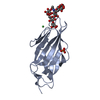 8bxyC 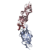 8by3C  2vcoS S: Starting model for refinement C: citing same article ( |
|---|---|
| Similar structure data |
-Links
 PDBj
PDBj- Assembly
| Deposited unit | 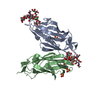
| |||||||||||||||||||||||||||||||||||||||||||||||||||||||||||||||||||||||||||||||||||||||||||||||||||||||||||||||||
|---|---|---|---|---|---|---|---|---|---|---|---|---|---|---|---|---|---|---|---|---|---|---|---|---|---|---|---|---|---|---|---|---|---|---|---|---|---|---|---|---|---|---|---|---|---|---|---|---|---|---|---|---|---|---|---|---|---|---|---|---|---|---|---|---|---|---|---|---|---|---|---|---|---|---|---|---|---|---|---|---|---|---|---|---|---|---|---|---|---|---|---|---|---|---|---|---|---|---|---|---|---|---|---|---|---|---|---|---|---|---|---|---|---|---|
| 1 |
| |||||||||||||||||||||||||||||||||||||||||||||||||||||||||||||||||||||||||||||||||||||||||||||||||||||||||||||||||
| Unit cell |
| |||||||||||||||||||||||||||||||||||||||||||||||||||||||||||||||||||||||||||||||||||||||||||||||||||||||||||||||||
| Components on special symmetry positions |
| |||||||||||||||||||||||||||||||||||||||||||||||||||||||||||||||||||||||||||||||||||||||||||||||||||||||||||||||||
| Noncrystallographic symmetry (NCS) | NCS domain:
NCS domain segments:
|
